Despite their benign nature, hemangioma has features of a clinically malignant course. Even pinpoint and small vascular tumors in newborns can show rapid growth, often reaching large sizes.
Despite the possibility of self-healing, its course still remains unpredictable.
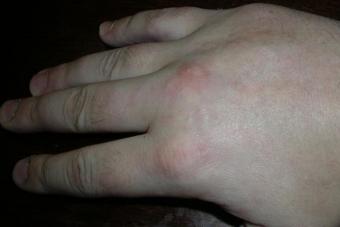
Simple hemangioma are red or blue-purple in color, located superficially, clearly demarcated, affecting the skin and a few millimeters of the subcutaneous fat layer, growing mainly to the sides. The surface of the hemangioma is smooth, less often - uneven, sometimes protruding slightly above the skin. When pressed, the hemangioma turns pale, but then again restore their color.
Cavernous hemangioma is located under the skin in the form of a limited node. It looks like a tumor-like formation, covered with unchanged or cyanotic skin at the top. When pressed, the hemangioma collapses and turns pale (due to the outflow of blood)
Combined hemangioma is a combination of superficial and subcutaneous hemangioma (simple and cavernous).
Mixed hemangioma consists of tumor cells emanating from blood vessels and other tissues. Appearance, color and consistency are determined by the tissues that make up the vascular tumor.
A feature of the course of some hemangiomas is their tendency to frequent ulceration and self-healing.
Hemangioma treatment
Surgical treatment of hemangioma is indicated for deeply located vascular tumors, when it is possible to remove the hemangioma entirely, within healthy tissues, without significant cosmetic damage. It is advisable to use the surgical method of treating hemangiomas in cases where the use of other methods of treatment seems to be obviously ineffective.
Hemangiomas of complex localizations are subject to radiation treatment, primarily tumors in areas where other methods of treatment cannot be used, for example, the orbital region. Radiation therapy is also indicated for simple hemangiomas of a large area.
Irradiation is carried out in separate fractions at intervals from 2-4 weeks to 2-6 months.
Only small, pinpoint hemangiomas are subject to diathermoelectrocoagulation in cases where the tumor is located in areas inaccessible to another method of treatment.
Bleeding can be considered an indication for electrocoagulation. Electrocoagulation of extensive and deep hemangiomas is not used.
Sclerosing treatment is indicated for small, deep-seated vascular tumors of complex localization, especially in the treatment of small cavernous and combined hemangiomas of the face and nasal tip.
For sclerotherapy, 70% alcohol or other drugs are used.
The disadvantages of sclerosing therapy are pain and duration of treatment. The advantage of injection therapy over other conservative treatment methods is its simplicity, which makes this method especially valuable.
One of the new methods of treating extensive hemangiomas of the external integument in children is hormonal treatment. Hormonal treatment is carried out with prednisolone. Hormone therapy is a fairly effective method of treating hemangiomas, however, with its high efficiency (98%), it is almost impossible to achieve the desired cosmetic result. This method perfectly stops the growth of a vascular tumor. Hormone therapy is no longer an independent, but an auxiliary method of treatment.
The use of low-temperature exposure. All simple hemangiomas of a small area, of any localization, are subject to cryogenic treatment.
The indication for the microwave cryogenic method of treatment is the presence of cavernous and combined hemangiomas with a pronounced subcutaneous part, more often with complex localization, not amenable or difficult to treat by other methods. The technique of microwave cryodestruction is quite simple, is carried out on an outpatient basis and does not require anesthesia. The hemangioma area is irradiated with a microwave field, followed by immediate cryodestruction.
In this article, we will consider what hemangioma is in newborns, how often it manifests itself, how hemangioma is diagnosed and treated in newborns, what are the dangers in the presence of hemangioma.
What is hemangioma in newborns
Hemangioma in newborns is a benign formation (tumor), in which changes occur in the structure of blood vessels (the structure is damaged). As a rule, the structure of the vessels in the hemangioma area is disturbed even before the birth of the child (during the embryonic period). Hemangiomas look, as a rule, as vascular points, or points that have merged into one spot. The color of a hemangioma can vary from pink to maroon (depending on which vessels are affected), it can be purple or vice versa, a bluish hue.
To the touch, the spot may be flat or bumpy (depending on the type of hemangioma). The size of the hemangioma can be completely different, from 1-2 mm to 15-20 cm or more. The shape can be approximately correct, with clear boundaries, or it can be completely wrong (like a spot with processes). It can be both outside and inside, under the skin or inside organs (different types of hemangiomas).
Superficial hemangiomas (flat, tuberous-flattened and tuberous-nodular, about them later) affect the skin 2-4 mm deep, there may be areas of deeper damage. Subcutaneous hemangiomas are blood-filled cavities and involve more than a few millimeters from the surface of the skin. Hemangioma can be either one spot or multiple spots.
Hemangioma, main signs
- When you press it with your finger, the hemangioma turns pale, and then gains color again.
- With, crying, the general tension of the child, the color (saturation) of the hemangioma may change, as a rule, the color becomes darker.
- The temperature of the skin around the hemangioma may be elevated.
- It can be in the form of a spot of any size.
- May be flat or convex, bumpy.
- Hemangioma consists of the same cells from which the inner surface of the vessels is built.
- It can increase in size (both in breadth and depth).
- This benign tumor has the fastest growth rate (if it grows).
Types of hemangiomas in newborns
In appearance and location, the following varieties are distinguished.
- flat hemangioma.
- Tuberous flattened hemangioma.
- Tuberous-nodular hemangioma.
These three types are superficial simple hemangiomas, are pink, red, or blue-burgundy in color, affect the outer part of the skin, and grow inside only a few millimeters. Their surface may be completely smooth, may partially protrude above the skin, may contain nodules.
- Cavernous hemangioma.
It can be both superficial and subcutaneous. Superficial is a cavity with blood and is visible on the surface of the skin. Subcutaneous - located under the skin, and looks like a tumor. It may be visible on the surface of the skin (a bluish tint to the skin), but it may not be visible, and the skin above it remains unchanged.
In addition to these forms, there are also combined forms of hemangiomas.
At what age does hemangioma most often appear?
Hemangioma can occur before the birth of a child, and is visible immediately after birth.
May appear at the age of 1 to 2 months (more often, during the first 2-3 weeks of life). But there are cases of manifestation of hemangiomas under the age of one year (these cases are rare).
Where is hemangioma in newborns
- Most often, hemangiomas affect the head area. They can be located on the scalp, on the mucous membrane of the mouth, eyes, nose, cheeks.
- Hemangiomas also often affect the mucous and skin parts of the genital organs.
- Hemangiomas can be located on any part of the body: arms, legs, abdomen, back, etc.
- Hemangiomas can affect internal organs, bones, soft tissues.
Hemangioma in newborns, causes
Until the end, the causes of hemangiomas in children are not known. There are several main reasons that doctors refer to, but they are quite vague, rather, they are theories.
I'll bring them.
- Unfavorable ecological situation, harmful factors in the environment during pregnancy.
- Past pregnancy diseases (and reactions to medications). As a rule, this is in the first 8 weeks of pregnancy.
- Hormonal changes in the body of a pregnant woman.
As you can see, there are no clear reasons. For some reason, it turns out that at a certain moment the cells of the inner surface of the vessels are placed in the wrong place, and are transformed into a benign tumor. As a rule, this occurs at the stage of pregnancy when the fetal cardiovascular system is being formed (from the third to the eighth, approximately, week of pregnancy).
- A common cause (except for the above) is considered to be tissue hypoxia, a lack of oxygen in the tissues.
Factors that make a hemangioma more likely to occur
Despite the fact that doctors have not yet identified clear causes of hemangiomas, there are factors in which the risk of occurrence increases. Let's bring them.
- Multiple pregnancy.
- Mother's age (over 37-38 years old).
- Low birth weight of the baby (less than 2900 g for a full-term pregnancy).
- Preterm pregnancy. Due to the fact that there is not enough surfactant (a substance for normal breathing) in the baby’s lungs, hypoxia may occur, and this can provoke the appearance of a hemangioma.
- A sharp increase in blood pressure (eclampsia) during pregnancy.
- Placental insufficiency, in which the function of the placenta, which is responsible for providing the fetus with oxygen, is impaired.
- Injuries during childbirth. Too fast, rapid labor, or vice versa, prolonged labor, cases of severe squeezing of the fetus. At this time, a state of local hypoxia occurs in places of compression, and this can provoke the appearance of a hemangioma.
- Smoking during pregnancy. This factor "works" in the same way as placental insufficiency. The fetus does not have enough oxygen, as the lungs of the mother are periodically filled with smoke.
Statistics of the appearance of hemangioma in newborns
Hemangioma is the most common benign tumor. On average, according to statistics, it can occur in every 10th child. More common in girls than boys. On average, there is 1 boy for every 3 girls with hemangioma.
The most common are simple (flat, tuberosity-flat and tuberosity-nodular) hemangiomas. This is about 70% of all cases. Hemangiomas of internal organs and bones are the least common. This is only 0.5% of all cases.
Hemangioma in newborns, treatment
There are cases when the treatment of hemangioma is carried out immediately, they are listed in the paragraph below. These cases, according to statistics, occupy about 10% of all cases. In all other cases, expectant management is recommended. You need to understand that small (especially superficial) hemangiomas quite often go away on their own, without treatment.
It is important to note the psychological moment in the occurrence of hemangioma. It may not apply to cases requiring urgent treatment, but at the same time it can be very psychologically traumatic for both the growing child and parents. The child will be "look askance", children may refuse to play with him or tease him. In these cases, a decision may be made to treat the hemangioma, even if it is not dangerous (and would probably disappear on its own with age).
Which doctor to observe and diagnose hemangioma
The following specialists may be needed to diagnose and monitor a child's hemangioma.
- Pediatrician.
- Pediatric surgeon.
- Children's dermatologist.
Depending on the location of the hemangioma, you may need to consult the following specialists.
- Children's ophthalmologist.
- Children's gynecologist.
- Children's ENT.
- Pediatric urologist.
- Children's dentist.
With complications in the development of hemangioma, consultations of the following specialists may be necessary.
- Oncologist (if malignancy is suspected).
- Infectionist (in case of infection of the hemangioma area).
- Hematologist, a specialist in blood diseases (for complications related to the circulatory system (for example, anemia or thrombocytopenia).
Diagnosis of hemangioma
To diagnose a hemangioma, the following procedures can be performed.
Medical examination. During the examination, the doctor (pediatric surgeon) finds out the history of the onset and development of the tumor. When it appeared, how it increased. In addition, he will measure its size, find out its structure, the nature of changes in the tumor under pressure.
Instrumental research. This group of studies is needed to identify internal hemangiomas (organs, tissues, bones), as well as when planning a surgical operation to remove the tumor. Instrumental studies may include:
- thermometry;
- thermography;
- ultrasonography;
- computed tomography;
- magnetic resonance imaging;
- angiography;
- biopsy.
Laboratory research(usually a complete blood count).
Consultations of related medical specialists, the main ones are listed above (gynecologist, ENT, etc.).
The process of development of hemangioma (phases)
As already mentioned, hemangioma appears, most often, in the first weeks of a child's life. After the appearance, it can grow intensively (up to about six months). The hemangioma, as a rule, reaches its maximum size by the year. Then regression often begins, resorption of the hemangioma, which can last up to 5-7 or 12 years.
In connection with the patterns of development, the phases of development of hemangioma are distinguished.
- First phase of development. The appearance of a small, relatively light, sometimes pinkish spot.
- Second phase of development. The spot begins to become more and more red, may resemble a scratch.
- Third phase of development. The hemangioma is rapidly growing in size (sometimes it doubles in a week).
- Fourth phase of development. The spot acquires a purple edge, the destruction of the subcutaneous layer begins and the germination of the hemangioma inside.
- Fifth phase of development. It is characterized by growth arrest (as a rule, by the year of the child, and up to 5-6 years it may increase slightly).
- Sixth phase of development. tumor regression. The surface becomes less bright, partly replaced by healthy skin, partly can be replaced by scar tissue. The complete disappearance of the tumor (generally without cosmetic defects) is observed in about 2 out of 10 cases.
How does a hemangioma go (disappear)
Many hemangiomas resolve without treatment. This is especially true for small flat hemangiomas. This course of events is called the spontaneous disappearance (spontaneous regression) of the hemangioma. As a rule, such age periods are characteristic of such spontaneous disappearance.
- Until the end of the first year of life.
- From one year to five or six years.
- Until the end of puberty.
With spontaneous disappearance, the following visual phenomena are observed.
- The appearance of light areas in the hemangioma, as a rule, first in the center, and then "spread" to the edges.
- A bulging hemangioma may become progressively flatter.
- Hemangioma may be replaced by scar tissue.
You need to understand that the hemangioma may disappear, but the cosmetic effect may be different. It can “leave without a trace”, or it can heal, and then it will be necessary to cosmetically correct the scar tissue. It may not go away completely, and then it will need to be corrected.
It should be noted that cavernous and combined hemangiomas themselves practically do not go away.
When is it necessary (mandatory) to treat hemangioma in newborns
- Hemangiomas in the mucosal area (eyes, nose).
- Hemangiomas in the labia or anus.
- Hemangiomas on the face.
- Sometimes hemangiomas in the neck.
- Hemangiomas that grow rapidly (about twice in 7-10 days).
- Hemangiomas on the inner surface of the cheeks and in the mouth (palate, tongue).
- Any hemangioma, anywhere, with signs of infection, bleeding, or necrosis.
- When signs of malignancy of the tumor appear.
Signs of malignant hemangioma
- A change in the quality of the superficial tissue of the tumor, a change in the usual structure of tissues, a sharp increase in breadth, height or depth. The appearance of ulcers, the appearance of peeling.
- Change in the usual consistency (composition) of the tumor. The appearance of denser, nodular areas.
- Change in color, the appearance of dark, black and brown areas.
- Changes in the skin around, inflammation, swelling, soreness, fever).
What is dangerous hemangioma in newborns
- Germination of hemangioma in the internal organs and their destruction.
- Damage and destruction of muscles and bones.
- Injury or compression of the spinal cord (may lead to paralysis).
- The appearance of ulcers and the penetration of infections into the hemangioma area.
- Malignancy.
- Cosmetic defects for life.
- (anemia) - a decrease in the concentration of hemoglobin in the blood, a decrease in the number of red blood cells.
- Thrombocytopenia is a condition associated with a reduced number of platelets, difficulty in stopping bleeding.
Removal of hemangioma in newborns
The following factors will be relevant to the selection of a specific removal method:
- Tumor size.
- Location of the hemangioma.
- A type of hemangioma.
It is possible to distinguish such groups of methods.
Physical removal methods
- Cryodestruction - freezing of hemangioma tissues (usually with liquid nitrogen). After freezing, the tissues are rejected. It is used for superficial or shallow located tumors.
- Laser irradiation is a modern and most aesthetically justified method of removing hemangioma. The risk of bleeding is minimal, since the blood in the vessels is sintered by the laser. The treated tissues do not form scars, which achieves the desired cosmetic effect.
- Sclerosing therapy - the method involves the introduction of special chemical solutions (for example, alcohol) into the hemangioma tissue, which cauterize the vessels and act as a coagulant.
- Electrocoagulation, - the impact on the hemangioma of high-frequency pulsed current. This method removes both superficial and internal hemangiomas, and can also prepare a hemangioma for surgery. A big plus of this method is that the vessels are cicatrized when exposed to electricity, the blood is sintered, and the risk of bleeding is lower.
- Close-focus X-ray therapy - local exposure to hemangioma tissue with X-rays. Often used as an additional method before surgery.
Surgical method of removal
It is used for small tumors, located in places where a scar will not have a cosmetic value. It is also used to remove internal tumors, with germination in organs and tissues. Surgical removal itself is often combined with other methods of influencing the tumor (drugs and physical methods discussed above).
Medical therapy
Drug exposure slows down the growth of hemangioma, can reduce its size. But medicines do not completely remove the hemangioma. Therefore, drug exposure is used as an additional method, for example, in preparation for the removal operation.
Dear moms and dads! If your child has a hemangioma, there are three main things to consider.
- On the one hand, do not miss the deterioration in its development, carefully observe it (both independently and with a specialist).
- On the other hand, it is important to be patient if the doctor advises so, and the hemangioma can go away on its own. You yourself understand that exposing the child to unnecessary stress during removal is not the best thing if there is a chance for complete disappearance without intervention.
- And on the third hand, it is also not worth exposing the child to negative psychological influences (if the age is already more than a year old, and the hemangioma is in a prominent place, and they literally “point the finger at the child”). If you need to wait at the cost of psychological discomfort and trauma to the child, then it is better not to wait, but to remove the hemangioma.
And, most importantly, do not self-medicate. This, although benign, is a tumor, and self-treatment is dangerous.
Health to your baby!
Hemangioma in children is a benign neoplasm with which a baby is usually born, or which appears in a child in the first month of life. According to statistics, hemangiomas are found in 10% of children. And if parents have the patience, knowledge and common sense not to try to treat a hemangioma, it will most likely go away on its own. Another thing is that most moms and dads almost never have enough: no patience, no knowledge, no sanity ...
What is a hemangioma
Not every neoplasm on the skin is rightly called a hemangioma. To understand exactly what's what, we will give clear signs of hemangioma:
- it is the most common benign tumor;
- it occurs only in children from birth to 1-2 months;
- hemangiomas occur in every 10th child on the planet;
- the exact causes of hemangiomas in newborns are still unknown to science;
- more often hemangiomas occur in girls than in boys (usually there is only 1 boy for every 3 girls with hemangiomas);
- hemangioma may be in the form of a flat spot, or may be convex, may increase in breadth, or may grow in depth;
- hemangioma in newborns can be of any size;
- the main feature that distinguishes hemangioma from any other neoplasms is that it consists of cells of the inner surface of blood vessels (endothelium);
- as a rule, if there are more than 3 hemangiomas on the surface of the skin, they are also on the internal organs of the child;
- in the vast majority of children, hemangioma resolves on its own;
The nature of the occurrence of hemangiomas in children
Alas, the exact causes and factors of hemangiomas in children are unknown to medical science. However, doctors are convinced that the nature of the occurrence of hemangiomas excludes heredity.
The mechanism of occurrence of hemangiomas in newborns is approximately as follows: the cells of the inner surface of the vessels (in scientific terms - the endothelium) at the stage of formation of the fetal cardiovascular system, due to some factors unknown to science, get "by mistake to the wrong address", and when the baby is born, they are transformed into a benign tumor that occurs on the skin, on the mucous membranes, and sometimes even on the internal organs of the child.
This tumor grows and develops for some time, and then, in most cases, self-destructs without any consequences.
Doctors believe that the risk of hemangiomas increases significantly if:
- multiple pregnancy (twins, triplets, etc.);
- mother at the time of birth is already over 38 years old;
- the child is born with low weight or prematurely;
- had eclampsia during pregnancy;
In premature babies, hemangiomas grow 2-3 times faster than in babies born on time.
We repeat that every tenth baby on the planet is born with hemangiomas (that is, with benign tumors consisting of endothelial cells). It often happens that not one or two tumors, but one or two dozen, are found on the baby's body.
But even in this case, you should not panic! Firstly, modern medicine has effective and safe methods for removing hemangiomas. And secondly - if these tumors are not touched, then by the age of 5-7 years of the child they will most likely disappear without a trace. Since in itself the life cycle of these neoplasms has its own natural ending.
Phases of existence of hemangiomas:
- Appearance, discovery(either it is already on the child's body at the time of his birth, or appears in the first 3-4 weeks after birth);
- Active growth phase(in all cases, tumor growth should end by 1 year):
- Growth arrest(the period when the hemangioma finally stops growing);
- Regression phase(the period during which the tumor gradually loses its size);
- Phase of involution(that is, the phase of biological regression and disappearance, which usually occurs at the age of the child 5, 7 or 9-10 years old);
The main rule: do not touch, but observe and fix
In the vast majority of cases, the hemangioma, formed in the “right” place, does not require any treatment, and even more so, surgical intervention. Nevertheless, it must be carefully and regularly monitored - with what dynamics it increases in size, where exactly the growth extends, whether it changes color, etc. Once a week or two it is necessary to show the pediatrician.
And the best thing is not just to observe the hemangioma in your baby, but also to take pictures. So that at the next appointment with a specialist doctor you can visually provide him with the entire dynamics of the development of hemangioma in the “before and after” format from the moment it appeared to the present day.
By the way, about doctors... It is very important that your child's hemangioma is observed by someone else. And a really good experienced specialist who encounters hemangiomas in newborns not a couple of times a year, that is, from time to time, but daily.
Remember: this is not a dermatologist, and not a cosmetologist, but none other than a pediatric surgeon. A referral to which you can always take from your attending pediatrician.
How and when does hemangioma in children go away
Hemangioma, of course, is not a pleasant thing, but fortunately it is not eternal. This benign neoplasm is distinguished, as we have already mentioned, by its excellent “patency” - that is, in most cases, with a policy of complete non-intervention, the hemangioma disappears on its own. In other words, the tumor simply gradually resolves, leaving almost no visible trace.
50% of all hemangiomas go away by the age of 5 years. 70% - by 7 years. The remaining 28-29% of children completely get rid of hemangiomas by the age of 9-10.
After the disappearance of the tumor, there is no risk of reappearance of hemangioma, or any negative consequences.
Cases in which removal of hemangiomas is indicated
An experienced and prudent doctor will never offer the parents of a baby who has hemangiomas to remove them without hesitation. In most cases, these tumors are only subject to regular monitoring and only. Because after 5, 7 or a maximum of 10 years they go away on their own.
Another thing is if the hemangiomas are located on the child's body in risky proximity to vital organs, or "behave", not corresponding to their nature. In these cases, the pediatric surgeon assesses the situation in detail and at each examination decides whether the hemangioma is to be removed now, or for some time it can simply be observed.
Situations in which hemangiomas are removed:
- 1 The formation of hemangioma on the mucous membrane. For example, if a hemangioma appears on the eye or on the larynx, or it grows into the ear cavity. The bottom line is that, entering the phase of active growth, hemangioma can irreversibly damage the organ. Or, if, for example, the tumor is located on the larynx, it can at some point block the access of air, which in itself is deadly.
- 2 Formation of a hemangioma in the immediate vicinity of physiological openings body (eyes, external auditory canals, mouth, nose, anus, genitals). The bottom line is that the growth of hemangiomas is unpredictable. Suddenly, the tumor can begin to actively grow in depth, touching or even blocking the natural physiological opening or passage. Which, in turn, can directly threaten the life of the baby.
- 3 The appearance and growth of a hemangioma in a place where it is easy to injure. For example, on the stomach, or on the side - just in the place where the belt of trousers or tights is usually located. Children, as a rule, simply pick off hemangiomas on the abdomen. Neoplasms in the area of the belt are injured with every inaccurate dressing and undressing. By itself, injury to a hemangioma is not dangerous - it will cover a little, like an ordinary wound, and will drag on. But repeated injury is always a reason for removal.
- 4 If the baby is already 1.5-2 years old, and the hemangioma continues to increase in size. Normally, by the year, the growth of any hemangioma should stop and gradually move into a phase of reverse development. If this does not happen, the tumor is removed.
- 5 If the child is already 10 years old, and the hemangioma has not disappeared on its own.
Removal of hemangioma - a strictly individual approach
If, for one reason or another, the doctor decides that the tumor should be removed, he selects the method on a strictly individual basis. When choosing, many factors are taken into account - for example, the size and location of the tumor.
The most common ways to remove hemangiomas:
- sclerosis;
- systemic hormone therapy;
- laser removal;
- cryodestruction.
Currently, in civilized countries, not a single pediatric surgeon approaches the removal of hemangiomas literally with a scalpel in his hands. Because modern medicine has long had many alternative, more gentle, ways to eliminate this problem.
For example, during cryodestruction of hemangioma, the scar, as a rule, does not remain. What is important if the tumor is located on the face or other open area. For deep-growing hemangiomas, a combination of cryodestruction with microwave radiation is usually used. First, the hemangioma is exposed to radiation, and then it is “frozen out” with liquid nitrogen.
In the event that the hemangioma "hidden" near the eye, the irradiation method is most often used. And if a hemangioma in a newborn occupies a large part of his body, then hormone therapy is used.
Often, a hemangioma in a newborn occurs not only on the surface of the skin, but also on the internal organs. Doctors noticed that if the baby has more than three hemangiomas on the skin, then most likely there are also tumors on the internal organs. In this case, constant monitoring is carried out not only visually, but also with the help of ultrasound. As a rule, internal hemangiomas are removed using hormonal therapy.

The issues of "self-passage" of infantile hemangiomas are actively discussed by both doctors and parents of patients. The information circulating in the media, near-scientific and scientific literature is very, very different. The numbers of self-regression (self-passage) of hemangiomas vary from 8 to 100%.
With this article, we want to answer most of the questions, based on the results of international studies and the data presented in the fundamental guide to vascular pathology Hemangiomas and Malformations. second edition. Edited by John B. Mulliken, Patricia E. Burrows, and Steven J. Fishman
Involution of infantile hemangiomas.
Growth of hemangiomas stops by the end of the first year of life. The following years, education develops in proportion to the growth of the child, and years later, a slow involution follows, the process of independent passage of the hemangioma.
The development of hemangioma corresponds to a certain pattern, graphically characterized by a dome-shaped graph (see figure). This curve characterizes the life (biological) cycle of infantile hemangiomas. The appearance and onset of development of hemangiomas is determined by the first month of a child's life, almost all children are born without any visible skin manifestations. The peak of development falls on the 4-5th month of a child's life, followed by a plateau period (growth stabilization) and after the first year there is a process of regression, withering of education. Although some hemangiomas continue to grow beyond 1 year of age (Bundling-Bennett et al. 2008).
The processes of active development (proliferation) and involution (process of passage) are not clear phases in the life cycle of infantile hemangiomas. The processes of apoptosis (the natural process of "disassembly" of pathological tissue) begin to prevail only after 1 year of life. But this process can be uneven in the same formation, for example, in some cases, involution processes can begin in the center of the formation, and proliferation (active growth) processes can actively take place on the periphery of the formation.
Immunohistochemical studies have shown that the final growth processes of infantile hemangiomas continue until the age of 4-5 years of the child (Mulliken and Glowacki 1982). The maximum process of apoptosis is reached at the age of 2 years (Raison et al. 1998).

Combination of involution processes in the center with active proliferation processes along the periphery of an infantile hemangioma.
One of the signs of the beginning of regression of an infantile hemangioma is a change in its color from bright crimson to lavender. The surface of the hemangioma is covered with a grayish shell, and upon closer examination, you can see the smallest white spots. The hemangioma becomes softer, less tense, the skin on the hemangioma becomes wrinkled. Hemangiomas become less hot, bleeding and ulceration cease to disturb the child. The processes of involution of infantile hemangioma begin, as a rule, from the middle of education and spread to the periphery.

The process of involution of an infantile hemangioma in the region of the upper third of the thigh on the right.
When a hemangioma appears, parents quite often note the child's anxiety, pain when in contact with the hemangioma. With the period of development of involution, hemangiomas become less painful, and the child is less capricious. Many parents notice that despite the passage of hemangiomas, they swell when crying, straining, with a rise in temperature and take on their former appearance when everything returns to normal. There is currently no reliable assessment of changes in blood flow in hemangiomas. The most reliable data on the decrease in blood flow in the hemangioma during an ultrasound study with Doppler sonography can be determined only at the age of 2-3 years, although in the majority, the pathological blood flow in the vessels supplying the hemangioma may persist even at an older age.

The process of involution of infantile hemangioma in the region of the right forearm. There are residual elements of hemangioma, telangiectasia, areas of healthy skin.
The process of involution continues from 1 year of life to 5-7 years. A change in the color of hemangiomas occurs by 5 years of age. Early clinical studies on the development of hemangiomas showed that complete resorption occurs in more than 50% of children by 5 years, and in more than 70% of children by 7 years, with continued improvement by 10-12 years. (Lister 1938, Pratt 1953, Simpson 1959, Bowers et al. 1960). Subsequent studies of children with hemangiomas showed that 80% of hemangiomas did not completely involute (“gone”) by 6 years of age and resulted in significant defects (Finn et al. 1983).
The involution of infantile hemangiomas is not affected by gender, race, tumor location, size, period of active growth, or morphological data (Bowers et al. 1960; Finn et al. 1983). The existing notion that large hemangiomas are less capable of regression than small tumors has been refuted by studies that have shown that tumor size does not affect the speed and extent of involution, and there is no relationship between the end result of involution and age (Simpson , 1959, Bowers et al. 1960).
The processes of involution are identical for all types of hemangiomas (superficial or deep). Prematurity does not affect the timing of involution. An interesting point of this study is that with hemangiomatosis (multiple hemangiomas), the process of involution occurs faster, by 2-3 years.
It has also been noted that involution processes occur most slowly in the region of the nose and lips (Bowers et al. 1960). A possible explanation for this circumstance can be considered that in the process of involution in this area it forms more fibro-adipose tissue. As a result, infantile hemangiomas may appear to heal more slowly in these areas.
In the case of continued growth of an infantile hemangioma in a child over 1 year old and no response to drug therapy, this formation requires a biopsy or complete removal for histological examination.
As a result of involution, practically healthy skin is restored in 50% of children with infantile hemangiomas (Finn et al. 1960). Quite often there is residual atrophy, telangiectasia (dilated capillaries, capillary "asterisks"), discolored skin. In the presence of a large, voluminous growth of an infantile hemangioma, as a result of involution, stretched, dough-like skin is formed.

The result of medical involution of infantile hemangioma in the back. As a result, a fairly large amount of fibrous and adipose tissue remained. Wrinkled, deformed and atrophic skin covered with superficial telangiectasias is determined. Veins show through the thinned areas of the skin.
After the involution of a convex tumor with clear boundaries, it is possible to show the draining veins through the skin, which may make this area appear cyanotic. If ulceration occurred during the active development of the hemangioma, then as a result, the ulceration site will become a pale scar, skin restoration in this area is impossible. An interesting observation is the propensity for acne or juvenile acne on the skin of an involuted hemangioma.
Bulging hemangiomas of any size usually result in a fibrofatty remnant. Deep infantile hemangiomas, without cutaneous manifestations, may completely regress without leaving any cosmetic cutaneous changes.
Hemangiomas in the scalp, as a result of active development, can injure the hair follicles, which can subsequently lead to reduced hair density in this area.
Periorbital hemangiomas often lead to proptosis, blepharoptosis, as well as an imbalance in the muscles of the eyeball.

The result of the growth of hemangioma in the orbit. There is a displacement of the eyeball.
Hemangiomas at the tip of the nose expand the lower lateral cartilages and leave a fatty residue, leading to a spherical enlargement of the tip of the nose.
Hemangiomas in the lip area often cause local hypertrophy (enlargement), erasure of the red border of the lip, sometimes leading to discoloration of the red border.

The result of the growth of hemangioma in the region of the upper lip. The shape is broken, the red border in the hemangioma area is not differentiated. Not only cosmetic, but also functional disorders are determined; it is difficult for a child to eat.
In most cases, the main problem that arises in a child, as a result of independent involution of an infantile hemangioma, is cosmetic defects of varying severity, requiring, subsequently, certain surgical or dermatological procedures to eliminate them. Another important factor is the social adaptation of the child in the children's team, the presence of cosmetic defects leads to problems in communication and attitude towards a child with hemangioma. In the near future, we will devote a separate article to this topic.
I thank Xenia Sofenko for help in translating.
Adapted translation of Mulliken and Young's Vascular Anomalies: Hemangiomas and Malformations
// November 16, 2014First of all, it must be taken into account that an online consultation cannot replace a full-fledged medical examination and dynamic monitoring by a doctor.
In this situation, the most important step is to conduct a differential diagnosis between hemangioma and vascular malformation, which determines the choice of the correct treatment tactics. The anamnesis of illness described by you: have noticed a swelling in 3,5 months; up to 8 months, the tumor increased in size; and from 8 to 11 months it practically did not change (only increased by 3 mm), in our opinion, it fits more into the classical version of the course of hemangioma: the tumor was detected after birth, there was a period of tumor growth, and now the size has stabilized. Unlike hemangiomas, vascular malformations, as congenital malformations of blood vessels, appear from birth and most often increase in proportion to the growth of the child (very slowly). However, the localization of the neoplasm in your son deep in the soft tissues makes it difficult to initially assess the size of the pathological process and does not exclude the presence of a vascular malformation. There are good laboratory tests that can detect hemangioma, but this diagnostic method is applicable only in the growth phase of the tumor. Given the different clinical course of hemangiomas and vascular malformations, dynamic observation in this case can be used as a method of differential diagnosis. The involution of hemangiomas occurs on average from one year of a child's life and lasts about 6 years. Being observed by your doctors, with regular ultrasound monitoring, you can evaluate changes in the volume of the neoplasm.
As for the clinical case described by Dr. Mikhail Valeryevich Zhitny, this is an extreme option that cannot prove the validity of the surgical treatment of all types of vascular anomalies. In this situation, there was no dynamic observation and I agree with my colleague that the treatment should have been carried out much earlier. However, we also have opposite clinical observations, when unjustified aggressive treatment of hemangiomas in children at an early age subsequently led to serious aesthetic defects, the correction of which required complex plastic and reconstructive surgeries.
I agree with my colleague that, until now, one of the main obstacles to the development of the field of medicine dealing with the study of anomalies in the development of blood vessels is confusion in terminology. The fact is that most often doctors confuse hemangiomas and angiodysplasias (vascular malformations), although their biological classification was proposed back in 1982 (Mulliken JB, Glowacki J. Hemangiomas and vascular malformations in infants and children: A classification based on endothelial characteristics Plast Reconstr Surg. 1982;69:412–422). If we talk about hemangiomas, then these are endothelial benign vascular tumors and their structure depends little on whether the tumor is located on the skin or under the skin. The division of hemangiomas into cavernous and capillary (classification by S.D. Ternovsky, 1959) in practice leads to confusion, since a doctor often associates a cavernous hemangioma with a vascular malformation, which leads to the choice of erroneous treatment tactics.