A leiomyoma is a medical term for a smooth muscle tumor. Most often it is benign, but it should be remembered that there is always a risk of its rebirth. It can form anywhere where there is smooth muscle - this is primarily the uterus, intestines and skin. Also leiomyoma may occur in the bladder or prostate gland.
Causes of Leiomyoma
The main causes of this disease on the skin are injuries and heredity. But the formation of a tumor in the gastrointestinal tract can occur against the background of numerous operations (opening the intestinal cavity, etc.), inflammation or unbalanced nutrition (if we are talking about excretory systems).
Also, risk factors are hormonal disorders, which may occur due to regular abortions or for other reasons.
Signs of the disease
It is difficult for a non-specialist to recognize leiomyoma, very often it proceeds without symptoms. The easiest way to do this is with a skin disease - red, red-violet or brown nodules the size of a pinhead or a little more form on the dermis. When they are pressed, rubbed or cooled, painful sensations occur.
Be sure to pay attention to any bleeding from the anus and heavy menstruation. Also, signs of leiomyoma can be regular pain in the pelvis, in the intestines, frequent urination, dysbacteriosis, feeling of a lump when swallowing, infertility, spontaneous miscarriages. But such symptoms appear at later stages. That is why doctors advise to undergo a full examination at least once every six months, because leiomyoma are best treated in the early stages of their development.
Leiomyoma treatment
First of all, if leiomyoma is suspected, the doctor prescribes an x-ray or ultrasound examination of internal organs (with the exception of skin diseases). Then if tumor detected and biopsied. You may also need to consult various specialists - a gynecologist, gastroenterologist, dermatologist, urologist and oncologist.
Once the diagnosis is confirmed, the tumor is usually removed. This can be done by cauterization (electrocoagulation) in the early stages, or by excision (surgical removal) if the mass is large. After surgery, the risk of recurrence is minimal.
There is no specific prophylaxis for leiomyoma. You should avoid abortions, injuries, hormonal disruptions, lead a healthy lifestyle and have regular check-ups with your doctor.
Leiomyoma (leiomyoma; from Greek leios - smooth and mys, myos - muscle) is a benign tumor originating from smooth muscle fibers.
Leiomyomas can occur in all organs where smooth muscle fibers are present, but are more common in the uterus, digestive tract, bladder, prostate, and skin. By origin, leiomyoma of the skin, esophagus, intestines are considered as dysontogenetic formations, uterine leiomyoma - as a consequence of endocrine disorders.
The tumor is round in shape, clearly delimited from the surrounding tissues; its consistency is dense, especially with a high content of connective tissue (leiomyofibroma). Leiomyoma nodes are often multiple, varying in size from microscopic to the diameter of the head of a full-term fetus and more; on the cut, it is pinkish, gray-white in color with a peculiar layered pattern due to the intersection of differently located muscle bundles. Microscopically, leiomyomas are built from muscle fibers that are somewhat larger than normal ones. Tumor cell nuclei are also relatively larger and richer in chromatin. In leiomyoma, muscle fibers form randomly arranged bundles, and sometimes they are placed concentrically around the vessels, in the form of muffs. Vessels are usually few; they are thin-walled, with a narrow lumen; rarely, leiomyomas contain a large number of dilated vessels (cavernous leiomyoma).
In long-term leiomyomas, as a result of circulatory disorders, dystrophic and atrophic changes in muscle fibers can be observed with their replacement by connective tissue; the latter can be subjected to hyalinosis, petrification, less often ossification. In addition, foci of necrosis, hemorrhages with the formation of cysts can occur in leiomyomas. Possible malignancy of leiomyoma (see Leiomyosarcoma). The treatment is surgical, the prognosis is favorable.
skin leiomyoma(synonym: myoma cutis, dermatomyoma) is usually a benign skin tumor originating from smooth muscle tissue. There are single leiomyomas of the skin, single leiomyomas of the genital organs (these leiomyomas have some structural features) and multiple leiomyomas of the skin. Histologically, all skin leiomyomas are characterized by interlacing of bundles of smooth muscle fibers with bundles of collagen tissue.
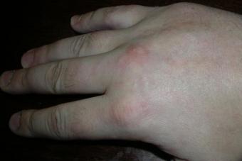
Solitary skin leiomyomas (synonymous with solitary angioleiomyomas) are formed from the smooth muscles of the venous wall, they are nodes that usually do not exceed 1 cm, less often 1.5 cm in diameter. They are located in the thickness of the dermis, protrude above the surrounding skin in the form of oval or rounded pinkish-yellow tumors, sometimes with a brown tint. The boundaries are clear (tumors are encapsulated), the consistency is dense. Quite often tumors are penetrated by blood vessels. On the skin of the face and extremities, more often the upper ones, groups of mobile tumors that are not interconnected can be observed. On palpation, there is a sharp soreness, but it can also occur spontaneously. Paroxysmal pains develop suddenly and also suddenly disappear after 1.5-2 hours. Pain and "tension" of the tumor may be aggravated by cold.
Single leiomyomas of the genital organs can develop on the scrotum, labia majora, in the area of the nipples of the mammary glands. These tumors are pinkish-yellowish in color, up to the size of a hazelnut, usually non-tense.
Multiple skin leiomyomas - no more than 0.5-0.7 cm in diameter, often tense, can be disseminated and in groups, often combined with uterine fibroids. Cases of illness of several family members are described, as well as the development of multiple skin leiomyomas at the site of injury.
Single genital leiomyomas and multiple skin leiomyomas are not encapsulated; the number of blood vessels in them is small, the amount of collagen is usually very significant; located in the dermis.
The course of skin leiomyoma is long, the prognosis is favorable, malignant degeneration is extremely rare. Treatment: surgical, electrocoagulation, application of carbonic acid snow.
Leiomyoma- a benign tumor of smooth muscle cells.
Epidemiology
Myogenic tumors account for about 10% of all soft tissue neoplasms of the skin. The ratio of smooth muscle tumors and neoplasms from striated muscle is 100:1. Pilar leiomyomas account for about 10% of all skin leiomyomas. Multiple forms are much more common than solitary ones. The ratio of women and men is 2:1, in familial cases - 8:1, in sporadic cases - 5:1.Classification
A leiomyoma that develops from the muscle that raises the hair (pilar leiomyoma).Dartoid, or genital leiomyoma, originating from the smooth muscles of the scrotal membrane, female external genitalia, or muscles that squeeze the nipple of the mammary glands.
Angioleiomyoma, which develops from the muscle elements of small vessels of the skin.
Modified classification of multiple skin leiomyomas according to E.I. Fadeeva (2002):
heredity: hereditary; sporadic (non-hereditary);
clinical variant: multiple isolated, focal, mixed, special forms (like Darier's disease, zosteriform variant, like neurofibromatosis).
Etiology and pathogenesis of leiomyoma
In multiple leiomyomas with an autosomal dominant type of inheritance, an association was found with some HLA-B8 haplotypes.
Clinical signs and symptoms
Multiple leiomyoma occurs at a young age, characterized by the appearance of small nodules of normal skin color, pink, red or other shades without subjective sensations. The nodules increase in size and number. The first elements appear on the limbs, less often on the back, chest, face. Pain sensations of varying degrees are present in almost all patients, usually paroxysmal in nature, lasting from several minutes to 1.5-2 hours.Solitary leiomyoma has the same appearance, but the elements are much larger.
Diagnosis is based on clinical signs and biopsy findings.The tumor node of the pilar leiomyoma is clearly delimited from the surrounding dermis and consists of intertwining thick bundles of smooth muscle fibers, between which there are narrow layers of connective tissue. When stained according to the Van Gieson method, the muscle bundles are stained yellow, and the connective tissue is stained red. A tumor that develops from diagonal muscles, without clear boundaries, has a similar structure, but the bundles of muscle fibers are somewhat thinner and lie more loosely. Between the muscle bundles in the sparse connective tissue are capillary-type vessels, sometimes with focal lymphohistiocytic infiltrates. Edema and dystrophic changes may be observed. The presence of a pronounced connective tissue stroma in painless tumors is probably one of the factors preventing excessive compression of nerve fibers during smooth muscle contraction.
Angioleiomyoma consists of a dense interweaving of bundles of thin and short fibers, located randomly in places, in places in the form of concentric structures or eddies. The tumor tissue contains many cells with elongated nuclei that are intensely stained with hematoxylin and eosin. Among these elements, many vessels are found with an indistinct muscular membrane, which directly passes into the tumor tissue, and therefore the vessels look like slits located between bundles of muscle fibers. Depending on the nature of the prevailing vascular structures, 4 main types of angioleiomyoma structure can be distinguished. The most common is angioleiomyoma of the arterial type, then venous and mixed, as well as poorly differentiated angioleiomyomas, in which a few vessels are determined, mainly with slit-like lumens. In some angioleiomyomas, one can see similarities with Barre-Masson's glomus angiomas. They are characterized by the presence of "epithelioid" cells that make up the bulk of the tumor. In later periods, various secondary changes can be detected in angio-leiomyomas in the form of a sharp expansion of blood vessels, proliferation of connective tissue, leading to sclerosis, hemorrhages, followed by the formation of hemosiderin.
Differential Diagnosis
Neurofibromatosis, leiomyosarcoma, neurinoma.General principles for the treatment of leiomyomas
Showing surgical excision, electroexcision of the solitary element.For relief of pain, a-blockers are shown in combination with calcium channel blockers: Nifedipine inside for a long time, 10 mg 3 r / day
+
Prazosin inside for a long time at 0.5 mg 3 r / day.
Forecast
With solitary tumors it is favorable, with multiple tumors it is relatively favorable.Leiomyoma of the skin is a benign tumor that develops from the smooth muscle elements of the skin. It occurs more often in men in young and middle age.
Synonym skin leiomyomas: dermatomyoma.
Allocate:
- multiple leiomyomas of the skin, the number of which sometimes reaches several hundred, developing from the smooth muscles of the skin;
- solitary, or dartoid, leiomyomas of the skin of the genital organs and smooth muscles of the nipple of the mammary gland;
- vascular leiomyomas, developing from the muscular membrane of the vessels of the skin.
pathological anatomy
At pathological anatomical examination, skin leiomyomas have the form of nodules and nodules of pinkish color, 1-3 cm in diameter (dartoid leiomyomas can reach 4-5 cm in diameter). Multiple and developing from small vessels leiomyomas often affect the skin of the upper extremities; leiomyomas emanating from the trailing arteries - the skin of the lower extremities with a favorite localization in the area of the joints of the lower leg and foot.
Microscopic examination shows that skin leiomyomas are built from bundles of smooth muscle fibers that intertwine in different directions. In dartoid tumors, muscle fibers are thickened, separated by uneven layers of fibrous tissue containing small vessels. Vascular leiomyomas of the skin are characterized by a large number of vessels of various sizes, resembling arteries and veins, or small vessels in the form of slits with indistinct muscle walls, directly passing into the tumor tissue.
Clinical manifestations
The clinical picture is characterized by pain on palpation and sensitivity of the tumor to a decrease in ambient temperature.
Diagnostics
The diagnosis is made on the basis of clinical and pathological findings.
Treatment
Treatment of skin leiomyoma is excision of the tumor within healthy tissues.
Forecast
The prognosis for skin leiomyoma is favorable. Relapses after surgery are extremely rare.
Big Medical Encyclopedia 1979
In some cases, tumors are benign, that is, they do not pose a particular danger at the moment, but over time they can either regenerate or begin to ulcerate. Leiomyoma is one of these tumors that predominantly affects the stomach.
Many patients leave the gastroenterologist's office with a diagnosis of gastric leiomyoma. At first glance, the term looks rather intimidating. But if you figure it out, then compared to malignant tumors, it will not be so dangerous. Leiomyoma is a tumor that is formed mainly from smooth muscle tissues. Most often it reaches 2 cm in diameter, but there are times when the size increases to five.
Such a tumor grows slowly, but, like all others, under the influence of certain factors, it can quickly begin to increase in size. The main feature of leiomyoma is that it has no effect on nearby organs, that is, all its negative effects stop only on the stomach. This happens because it is formed from its own tissues, without cell degeneration, that is, in fact, it is not alien.
If you do not contact a specialist in time when the very first symptoms of gastric leiomyoma appear, it is possible that it will degenerate into leiomyosarcoma, which is already a malignant tumor and progresses very quickly.
Reasons for the formation of a tumor

In the body, a failure never just happens, and in order for even benign tumors to begin to appear, negative factors must influence. The main reasons for the formation of leiomyoma in the stomach include:
- Wrong nutrition. As a result of the fact that a person eats improperly, mainly fatty and spicy foods, the mucous membrane begins to break down, and no longer copes with the task. In this case, a general weakening of the body occurs, due to which the smooth muscle cells on the walls of the stomach begin to grow rapidly.
- Radiation. This usually includes both radiation and electromagnetic radiation, which literally affects a person every day. A huge amount of equipment, access systems - all this negatively affects the cells. People who often fly on airplanes are most susceptible to the development of the tumor, as they have to pass through arches with x-rays several times.
- Polluted air, which also pollutes the lungs, and after them all the organs do not receive the amount of oxygen that they so need. In addition, particles of chemicals enter the body, which are deposited on the bronchi.
- Bacteria and viruses that enter the body so quickly that a person does not always have time to react and start fighting them. And it’s good if their presence manifests itself with obvious symptoms, but most often they destroy the body asymptomatically. So, the stomach is negatively affected by a bacterium which, according to scientists, causes such dangerous diseases as stomach ulcers, gastritis, and sometimes cancer.
- Injuries during which internal organs were damaged and the stomach was affected
- Weak immunity, as a result of which the body is most susceptible to negative factors. Due to reduced immunity, cells do not react in any way to changes
- Problems with hormones. Hormones are especially dangerous, namely when their level changes dramatically and becomes either too high or too low. Under the action of hormones, tumors can form, cells can be reborn, etc.
- Inflammatory process in the stomach, namely inflammation of the mucous membrane
- Smoking and. During smoking, all cells are literally saturated with nicotine, which can kill them. But alcohol gradually burns the mucous
- Heredity. A special role is played by heredity. If there are people in the family (close relatives) who suffered from gastric leiomyoma, then the likelihood that it will develop increases several times
- Stress. Under the influence of stress, irreparable changes occur in the body, which most often affect the stomach and nervous system. Constant nervous tension is sometimes more dangerous than radiation
Usually, gastric leiomyoma develops in people over the age of 50, since it is from this moment that the body becomes less resilient, and the cells gradually age.
Symptoms of gastric leiomyoma

The success of treatment will depend on how quickly a person notices the first manifestations of gastric leiomyoma. That is why, each person should know what symptoms it will manifest. In most cases, the disease is asymptomatic, since the tumor does not have any effect either on the stomach itself or on other organs. But over time, ulcers may appear on it, which are dangerous for the onset of bleeding. Symptoms of leiomyoma include:
- Weakness, as well as dizziness, which appear when the tumor begins to bleed. Blood loss always leads to such symptoms, but in this case, the bleeding will not be noticeable to the person.
- Decrease in body weight. Due to the fact that the gastric mucosa is destroyed by the influence of the formed tumor, especially when it begins to ulcerate, a person loses weight. This happens for one reason - nutrients are not able to be absorbed normally when there is blood in the stomach, and the mucous membrane does not perform its intended functions.
- Anemia. Sometimes, when taking a complete blood count, the doctor may notice a slight anemia, which is not dangerous. But when leukocytes are present in the blood, and hemoglobin is much lowered, then this is a clear sign
- Heartburn, which appears due to excessive injection of gastric juice from the esophagus. This happens because when ulcers appear on the tumor, the muscles weaken
- Painful sensations, mainly this (which happens when a person wants to eat) and at night, especially after a late meal. It is not uncommon for people with gastric leiomyoma to wake up at night with pain in the stomach area. And not always painkillers relieve pain
- Chair painted dark. Dark-colored stools are one of the very first signs that bleeding has begun in the intestines or stomach.
- Pale skin that becomes so due to the loss of iron in the body, as well as due to constant weakness
- Fatigue, even when a person is resting as much as doctors advise
As a rule, all the above symptoms appear only when the tumor has grown so much in size that it begins to interfere with the normal functioning of the stomach. And the main danger of leiomyoma due to its asymptomatic course will be the possibility of ulcers and the onset of bleeding, which can threaten a person's life.
Diagnosis of the disease

The Ministry of Health in our country recommends that every resident undergo an annual medical examination so that problems can be identified at an early stage and treated in a timely manner. But it is not always on the list of main doctors, and even if he is there, few people will simply prescribe a procedure for a person to examine the state of the gastrointestinal tract.
If a person suffers, then at least once every two years he will do this procedure in order to protect himself from the appearance of an ulcer (and for control). And it is during the FSH that most specialists detect leiomyomas, and by chance, because, as mentioned above, at the very beginning it does not manifest itself with any symptoms. If the person nevertheless began to be tormented by symptoms, then the attending physician will carry out the following procedures, after which an accurate diagnosis will be made:
- Collection of anamnesis. During the conversation, the doctor will have to find out what diseases the next of kin had, who died of what, what complaints the patient currently has, what lifestyle he leads, etc. Many questions will be asked that will help the specialist to learn more about the person.
- Examination of the patient. The doctor will examine his skin, stomach, check the perception of pain, etc.
- Passing tests, among which there will be a general analysis of blood and urine, as well as feces. According to the results of the coprogram, the doctor will be able to know for sure whether there is bleeding in the gastrointestinal tract or not
- Carrying out during which the doctor will examine the condition of the mucous membrane of the stomach, esophagus, etc. and if necessary, take a biopsy
- Getting tested for the presence of bacteria such as Some prefer to do a breath test, while others rely more on blood tests
- Ultrasound examination, during which the specialist can accurately determine whether there is a tumor or not. And also find out its approximate size and location.
- Computed tomography, the main purpose of which will be to identify the origin of the tumor
After the doctor has received all the results after the above procedures, he can accurately diagnose. And even if his suspicions of the presence of leiomyoma are confirmed, then with the right treatment, the prognosis will be good.
Leiomyoma treatment

Treatment of leiomyoma will directly depend on what size it has reached. Human susceptibility also plays a huge role. Some people live normally with a three-centimeter leiomyoma, while others experience severe discomfort with a one-centimeter one. Basically, experts offer patients, since conservative methods are not able to have the desired effect. In the event that the tumor is small, it can be removed using an endoscope. But if its size is large, then you will have to perform an abdominal operation, which is much more dangerous.
The choice of treatment method also depends on the patient's response to medication. There are times when a person cannot tolerate anesthesia. Then endoscopy is performed and the tumor is frozen. After removal of the tumor (by any means), the patient is prescribed medication, the main purpose of which is to reduce the inflammatory process and reduce the risk of complications. Usually, patients are prescribed drugs that will reduce the production of hydrochloric acid, as it can further damage an already damaged surface, as well as antibiotics. discharged when the presence of the Helicobacter bacterium was detected.
Leiomyoma of the stomach, despite the fact that it refers to benign tumors, can cause great discomfort to a person. Many patients, knowing that it does not pass to other organs, treat it negligently, thereby increasing the likelihood of complications (ulcers, bleeding, degeneration). But this is a big mistake, since timely treatment eliminates a long recovery period, and reduces the likelihood of abdominal surgery.
You can learn more about stomach cancer from the thematic video: