Registration for a consultation with leading specialists in this section of medicine is carried out by phone: 8-918-55-44-698
Suppuration of a postoperative wound is an article that will tell about the features of the development of the process of wound suppuration after surgery.
Determination of the process of suppuration of the wound
Any surgical intervention is accompanied by suturing at the site of the incision. This is a classic way to complete the operation, which is practiced in most medical institutions to this day, despite the existence of laser treatment. It is worth recognizing that the operation is sometimes the most acceptable way to save a person's life. However, various complications that can occur in the postoperative period are of great danger, and often they are associated with suppuration of the suture at the wound site. This is a common occurrence that can be stopped under certain circumstances and timely detection. So, what can be associated with the process of suppuration?
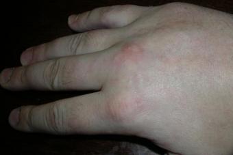
Note that after surgery, a kind of failure occurs in the patient's body, because the process is a stress for the functioning of the immune systems. Weakness leads to frequent complications, one of which is purulent inflammation of the wound. It is worth saying that the appearance of pus is always a signal that indicates the presence of an inflammatory focus, in which the patient often experiences the following symptoms: fever, chills, general weakness, burning at the site of wound stitching. The skin around the site of suppuration becomes tight, redness and swelling are noticeably manifested. The development of swelling indicates the beginning of the process of suture infiltration.
Suppuration of the postoperative wound
Causes of suppuration of the postoperative wound
The fact is that in most cases the wound is vulnerable to the penetration of dangerous microorganisms and infections. The ingress of viral bacteria can cause wound intoxication in a short time, the rapid multiplication of the infection is the cause of the melting of the affected tissues and the main feature of suppuration. Despite the use of antibacterial measures in the postoperative period, the likelihood of purulent inflammation is high. Usually the first signs are observed within 6-8 hours after the completion of the surgical intervention.
This is a natural process that is associated with an immune failure, which means that the treatment of purulent lesions of the wound should be carried out by combined methods. The most important thing in preventing the pathological process is the use of antibiotics and antiseptics to care for skin inflammation. It is necessary to stop the purulent spread and increase the patient's immunity. To do this, the attending physician performs a second operation, during which the affected tissues are opened and a special treatment of the wound is performed. It is possible to use the drainage method, especially in the case of closed suppuration. Subsequently, upon successful completion of the treatment of the lesion, the wound is sutured a second time.
Suppuration Wounds- Suppuration of the surgical wound ( suppuration of the postoperative wound, or suppuration of the seam) today has a number of features. First of all, the frequency of this complication has increased (according to many authors, from 1 to 15% or more - A. I. Gnatyshak and L. R. Kryshtalskaya, 1967; B. V. Petrovsky, 1971; V. A. Proskurov, 1974; Altemeier , 1970; Bruun, 1970; Grun 1974; Brock, 1975, and others; 5.4% of all operations in our observations). The increase in the number of suppurations, in addition to the general causes of the growth of nosocomial infection, can be explained by a number of factors:
- the initial state of the patient and his unsatisfactory defensive reaction;
- complications that developed during the operation and due to errors in operational equipment;
- infection of the wound during or after surgery.
Thus, after operations with cardiopulmonary bypass, the wounds have some features due to a mild inflammatory reaction and a slowdown in regeneration. A similar course of the wound process was noted during organ transplantation with the use of immunosuppressants, after a severe injury, in patients with congenital or acquired immunological deficiency. These circumstances caused them to have a high frequency of festering wounds.
According to the clinical course, patients with suppuration of wounds can be divided into three groups. In patients of the first group, local signs were expressed. General well-being did not suffer significantly. Only a temperature reaction was noted. The outcome was usually good. In the second group, a more severe general course was noted, accompanied by severe intoxication, secondary exhaustion, and prolonged healing. In patients of the third group, the suppuration of the wound progressed, the process spread to the surrounding tissues, peritonitis, mediastinitis, empyema of the pleural cavity, pneumonia, sepsis and other complications accompanied by septicemia, septic shock often joined. They were preceded by some degree of unresponsiveness. The prognosis has always been serious.
Suppuration of the wound usually proceeded with the second wave of temperature increase (on the 5th - 8th day with staphylococcus aureus, on the 3rd - 5th day - with Pseudomonas aeruginosa). Prolonged fever was more often observed, starting from the first postoperative day. Local signs of inflammation were somewhat delayed in time and were detected on the 7th - 8th day with staphylococcus aureus, on the 3rd - 4th day with Pseudomonas aeruginosa. Most patients, even before the appearance of local phenomena, noted a deterioration in well-being, pain in the wound, fever, sometimes chills, tachycardia, and shortness of breath. The temperature rose to 38°C and above. On examination and palpation, it was possible to detect pastosity and infiltration of the edges of the wound, in some cases areas of hyperemia and pain. Sometimes there was leakage of pus between the seams. After the sutures were removed, the edges easily diverged, edematous subcutaneous fat covered with a gray coating was exposed, a cloudy hemorrhagic fluid or pus was released.
In cases of wound infection caused by Pseudomonas aeruginosa, fibrinous-purulent inflammation was superficial, the pus was thick and viscous at first. On the 3rd - 4th day after dilution of the edges of the wound, the nature of the discharge began to change. The pus became more liquid, its color acquired a characteristic greenish-yellow hue, which is associated with the formation of a blue-green pigment - pyocyanin, which is released only under aerobic conditions. Therefore, the blue-green color of the dressings, especially their surface layers, is a very characteristic sign for a local Pseudomonas aeruginosa infection. Flaccid, pale granulations bled easily. There was a specific smell, which was sometimes noted from the first day.
When determining the pH of purulent wounds using universal indicator paper, it was found that Pseudomonas aeruginosa infection gives an alkaline reaction (pH 8.5 - 9.0), with staphylococcal suppuration, the reaction is slightly acidic or neutral (pH 6.8 - 7.0).
Thus, the following signs are characteristic for suppuration of a wound of Pseudomonas aeruginosa etiology: 1) staining of the surface layers of the dressing 1–2 days after dressing in a blue-green color; 2) copious liquid purulent discharge of blue-green color with a specific odor; 3) flaccid pale, easily bleeding granulations with significant edema and swelling of the wound edges; 4) fluorescence in the case of irradiation with long-wavelength rays in a darkened room; 5) alkaline reaction of the wound (pH over 8.5).
In a combination of several pathogens, Pseudomonas aeruginosa helps to get the predominance of the use of antibiotics, to which it remains the most resistant.
Morphological changes in most cases of suppuration of wounds were of the same type. The postoperative wound on the chest was a gaping hole with necrotic edges saturated with pus, sometimes with exposed ribs and scapula. The spread of the process to the surrounding tissues led to chondritis or osteomyelitis of the rib. In some cases, the infiltrate extended to the diaphragm. Quite often there was a communication with a pleural cavity, the empyema of a pleura developed. With median access, fibrinous-purulent inflammation passed to the anterior mediastinum, penetrating in some cases into deeper tissues and giving a picture of purulent mediastinitis, pericarditis, and sometimes osteomyelitis of the sternum. Suppuration of the postoperative wound of the anterior abdominal wall, which spread beyond the aponeurosis, could lead to communication with the abdominal cavity, peritonitis, and eventration.
read also
Article content: classList.toggle()">expand
Any surgical intervention, for whatever reason it is not performed, inflicts a wound on the patient, which then requires care until the moment of healing.
Quite often, unfortunately, in the process of restoring damaged tissues, various complications arise, the most common of which is suppuration. This happens regardless of how carefully and correctly the operation was performed, even after the perfect performance of all actions, the postoperative wound may begin to fester.
Causes of suppuration of the postoperative wound
Most often, the appearance of suppuration of postoperative wounds occurs due to:
Seam processing and dressing
Treatment of sutures after surgery is carried out at each with the help of antiseptic solutions and special preparations.
Before starting the dressing procedure, wash your hands thoroughly with soap and water.(it is recommended to do this up to the elbow), dry them with a paper towel and put on gloves. After that, you need to remove the applied dirty bandage. If the gauze has dried up in some places to the incision site, you should not tear it off, you just need to moisten the bandage with hydrogen peroxide in these places and wait a bit.
After removing the bandage, gloves must be changed or thoroughly washed and treated with a disinfectant solution. The seams and the tissue incision line should be shed or with a solution, blot with a sterile napkin and allow the skin to dry. If there is no suppuration, and blood does not ooze anywhere, then it is possible to treat the wound and the surface of the skin around it, as well as the sutures of the usual one, applying it in a thin layer once a day when changing the bandage.
If the postoperative injury has not yet dragged on, it is necessary after treatment to apply brilliant green only to the skin area around the incision line, and an ointment should be applied to the wound itself to prevent suppuration or eliminate it when inflammation has already begun.
An important point is that when treating wounds after operations and changing dressings, in no case should the scabs formed and the existing whitish coating be torn off.
Scabs and plaque formation indicate that the process of formation of new tissues and epithelium has already begun at the site of damage. An attempt to remove the scab and such a plaque hello to the formation of serious scars in the future.
Removal of stitches is carried out, as a rule, in the period from the 7th to the 14th day after the operation., which depends on the scale of the section and its complexity. The procedure is carried out without any anesthesia, since it causes pain to patients only in rare cases. Before and after the removal of sutures, the skin and the incision site are treated with antiseptics.
Treatment of inflammation
If signs of suppuration of the postoperative wound appear, it is necessary to start its treatment as soon as possible. Treatment of such a wound is carried out according to the same scheme as any other purulent wound and consists in frequent dressing changes with proper treatment with antiseptics, disinfectants and anti-inflammatory drugs.
The action of modern ointments is long-lasting, and the effects are pronounced, which allows you to heal postoperative wounds much faster and eliminate inflammatory processes, with virtually no side effects. Such indicators of many drugs make it possible to use them for the treatment of wounds and for a long time, if necessary.
 Application has many advantages. In particular, the ointment has a fairly thick but soft structure, which allows it to be applied to any part of the body without fear of dripping (unlike liquid preparations). The special formula of such agents allows them to quickly penetrate deep into damaged tissues, while creating a protective film on the surface of the wound.
The use of ointments is safer than injections or oral antibiotics, since ointments have only a local effect, without creating a systemic effect.
Ointment for the elimination of suppuration of postoperative wounds and the treatment of other purulent wounds should solve certain problems:
- Fight infection inside the resulting wound.
- Contribute to the removal of dead tissue and cleansing of purulent formations.
- Eliminate the inflammatory process, stopping its development.
- Do not create obstacles for the exit of pus.
- Protect the wound from the penetration of harmful microorganisms into it.
Similar articles 
The first stage of wound healing after surgery usually begins on the third day. During this period, you can use water-based ointments that promote faster healing of damage, eliminate inflammation, prevent the penetration of infection or suppress its development. These drugs include ointments:, Sulfamekol, Ichthyol, Dioxin, Zinc.
Ointments that activate regeneration processes and eliminate bacterial infections should be applied to the cleaned surface of postoperative injuries for faster recovery of injured tissues.
For the treatment of postoperative sutures, they are used because they contribute to the formation of higher quality granulation. Most often, in this group of drugs, ointments based on Tetracycline and Gentamicin are used.
You can also use universal ointments with a combined composition. Such agents are highly effective in eliminating the inflammatory process and accelerating wound healing. The drugs of this group include Vishnevsky ointment, Oxycyclozol, Solcoseryl, Levomethoxin.
Folk remedies
The treatment of sutures after operations with traditional medicine can significantly speed up the process of restoring injured tissues and avoid many complications. Traditional medicine has a wide variety of recipes.
The most effective folk remedies for the treatment of wounds after surgery:

Complications and consequences
The main complication after surgery is wound suppuration, which must be dealt with by all means.
Often, after the stitches are removed and the patient is discharged home, the inflammatory process begins again and re-suppuration occurs. This happens during secondary infection of a protracted wound, for example, in cases where a person begins to peel off the crusts formed along the incision line, thereby injuring new tissues. With such actions, harmful microorganisms can enter small wounds and cause a new inflammatory process.
Secondary infection with an infection or the spread of an existing one often occurs due to improper and insufficient suture treatments, when the patient neglects the timely change of dressings and does not use prescribed remedies.
After being discharged home, special attention should be paid to the condition of the sutures and the resulting scar. If a pronounced reddening of the skin, swelling, swelling of tissues, new purulent formations appear around it, you should immediately consult a doctor.
Suppuration of the Wound - Suppuration of the surgical wound (suppuration of the postoperative wound, or suppuration of the seam) today has a number of features. First of all, the frequency of this complication has increased (according to many authors, from 1 to 15% or more - A. I. Gnatyshak and L. R. Kryshtalskaya, 1967; B. V. Petrovsky, 1971; V. A. Proskurov, 1974; Altemeier , 1970; Bruun, 1970; Grun 1974; Brock, 1975, and others; 5.4% of all operations in our observations). The increase in the number of suppurations, in addition to the general causes of the growth of nosocomial infection, can be explained by a number of factors:
the initial state of the patient and his unsatisfactory defensive reaction;
complications that developed during the operation and due to errors in operational equipment;
infection of the wound during or after surgery.
Depending on the localization of suppuration, various variants of the clinical course were noted. On the chest, the purulent process usually proceeds more severely than on the abdominal wall or limbs. A particularly severe clinical course was observed with wound suppuration after operations with cardiopulmonary bypass. In this group of patients, the reactivity and immunological properties of the body change significantly. The inflammatory reaction slows down, becomes inferior, all reparative processes are disrupted. In this regard, there was often a divergence of the seams and rapid infection of wounds, the phenomena of hemorrhagic diathesis (in the form of multiple small hematomas along the wound). The growth of granulations and healing slowed down significantly. The delay in regenerative processes in wounds after operations with cardiopulmonary bypass led to a prolongation of their healing time. Histological examination of the wound edges showed a sharp decrease in the number of leukocytes and histiocytes. Fibroblasts and fibers of fibrous tissue changed pathologically: hypertrophied fibroblasts and thickened fibers appeared. Damage to the vascular wall, areas of hemorrhage, and hematomas were also observed. The wound surfaces were covered with a gray coating, emitted a putrid odor.
Thus, after operations with cardiopulmonary bypass, the wounds have some features due to a mild inflammatory reaction and a slowdown in regeneration. A similar course of the wound process was noted during organ transplantation with the use of immunosuppressants, after a severe injury, in patients with congenital or acquired immunological deficiency. These circumstances caused them to have a high frequency of festering wounds.
According to the clinical course, patients with suppuration of wounds can be divided into three groups. In patients of the first group, local signs were expressed. General well-being did not suffer significantly. Only a temperature reaction was noted. The outcome was usually good. In the second group, a more severe general course was noted, accompanied by severe intoxication, secondary exhaustion, and prolonged healing. In patients of the third group, the suppuration of the wound progressed, the process spread to the surrounding tissues, peritonitis, mediastinitis, empyema of the pleural cavity, pneumonia, sepsis and other complications accompanied by septicemia, septic shock often joined. They were preceded by some degree of unresponsiveness. The prognosis has always been serious.
Suppuration of the wound usually proceeded with the second wave of temperature increase (on the 5th - 8th day with staphylococcus aureus, on the 3rd - 5th day - with Pseudomonas aeruginosa). Prolonged fever was more often observed, starting from the first postoperative day. Local signs of inflammation were somewhat delayed in time and were detected on the 7th - 8th day with staphylococcus aureus, on the 3rd - 4th day with Pseudomonas aeruginosa. Most patients, even before the appearance of local phenomena, noted a deterioration in well-being, pain in the wound, fever, sometimes chills, tachycardia, and shortness of breath. The temperature rose to 38°C and above. On examination and palpation, it was possible to detect pastosity and infiltration of the edges of the wound, in some cases areas of hyperemia and pain. Sometimes there was leakage of pus between the seams. After the sutures were removed, the edges easily diverged, edematous subcutaneous fat covered with a gray coating was exposed, a cloudy hemorrhagic fluid or pus was released.
In cases of wound infection caused by Pseudomonas aeruginosa, fibrinous-purulent inflammation was superficial, the pus was thick and viscous at first. On the 3rd - 4th day after dilution of the edges of the wound, the nature of the discharge began to change. The pus became more liquid, its color acquired a characteristic greenish-yellow hue, which is associated with the formation of a blue-green pigment - pyocyanin, which is released only under aerobic conditions. Therefore, the blue-green color of the dressings, especially their surface layers, is a very characteristic sign for a local Pseudomonas aeruginosa infection. Flaccid, pale granulations bled easily. There was a specific smell, which was sometimes noted from the first day.
When determining the pH of purulent wounds using universal indicator paper, it was found that Pseudomonas aeruginosa infection gives an alkaline reaction (pH 8.5 - 9.0), with staphylococcal suppuration, the reaction is slightly acidic or neutral (pH 6.8 - 7.0).
Thus, the following signs are characteristic for suppuration of a wound of Pseudomonas aeruginosa etiology: 1) staining of the surface layers of the dressing 1–2 days after dressing in a blue-green color; 2) copious liquid purulent discharge of blue-green color with a specific odor; 3) flaccid pale, easily bleeding granulations with significant edema and swelling of the wound edges; 4) fluorescence in the case of irradiation with long-wavelength rays in a darkened room; 5) alkaline reaction of the wound (pH over 8.5).
In a combination of several pathogens, Pseudomonas aeruginosa helps to get the predominance of the use of antibiotics, to which it remains the most resistant.
Morphological changes in most cases of suppuration of wounds were of the same type. The postoperative wound on the chest was a gaping hole with necrotic edges saturated with pus, sometimes with exposed ribs and scapula. The spread of the process to the surrounding tissues led to chondritis or osteomyelitis of the rib. In some cases, the infiltrate extended to the diaphragm. Quite often there was a communication with a pleural cavity, the empyema of a pleura developed. With median access, fibrinous-purulent inflammation passed to the anterior mediastinum, penetrating in some cases into deeper tissues and giving a picture of purulent mediastinitis, pericarditis, and sometimes osteomyelitis of the sternum. Suppuration of the postoperative wound of the anterior abdominal wall, which spread beyond the aponeurosis, could lead to communication with the abdominal cavity, peritonitis, and eventration.
134. How is IVL (artificial ventilation of the lungs) carried out "from mouth to mouth", "from mouth to nose" with the help of the "AMBU" apparatus?
Artificial lung ventilation
I Artificial lung ventilation
provides gas exchange between the surrounding air (or a specially selected mixture of gases) and the alveoli of the lungs.
Modern methods of artificial lung ventilation (ALV) can be divided into simple and hardware. Simple methods are usually used in emergency situations: in the absence of spontaneous breathing (apnea), with acutely developed respiratory rhythm disturbance, its pathological rhythm, agonal type breathing: with an increase in respiration of more than 40 per 1 min, if this is not associated with hyperthermia (body temperature is higher 38.5°) or severe unresolved hypovolemia; with increasing hypoxemia and (or) hypercapnia, if they do not disappear after anesthesia, restoration of airway patency, oxygen therapy, elimination of a life-threatening level of hypovolemia and gross metabolic disorders. Simple methods primarily include expiratory methods of mechanical ventilation (artificial respiration) from mouth to mouth and from mouth to nose. In this case, the head of the patient or the victim must necessarily be in the position of maximum occipital extension (Fig. 1) to prevent retraction of the tongue and ensure airway patency; the root of the tongue and the epiglottis are displaced anteriorly and open the entrance to the larynx (Fig. 2). The caregiver stands on the side of the patient, with one hand compresses the wings of his nose, tilting his head back, with the other hand slightly opens his mouth by the chin. Having taken a deep breath, he presses his lips tightly to the patient's mouth (Fig. 3) and makes a sharp energetic exhalation, after which he takes his head to the side. The exhalation of the patient occurs passively due to the elasticity of the lungs and chest. It is desirable that the assisting person's mouth be insulated with a gauze pad or a piece of bandage, but not with a dense cloth. During mechanical ventilation from mouth to nose, air is blown into the nasal passages of the patient (Fig. 4). At the same time, his mouth is closed, pressing the lower jaw to the upper one and trying to pull his chin up. Air blowing is usually carried out with a frequency of 20-25 per 1 min; when combining mechanical ventilation with heart massage (see Resuscitation) - with a frequency of 12-15 in 1 min. Carrying out simple mechanical ventilation is greatly facilitated by the introduction of an S-shaped air duct into the oral cavity of the patient, the use of Ruben's bag ("Ambu", RDA-1) or RPA-1 fur through the oral mask. In this case, it is necessary to ensure the patency of the respiratory tract and tightly press the mask to the face of the patient.
Hardware methods (with the help of special respirators) are used if necessary for long-term ventilation (from several hours to several months and even years). In the USSR, the most common are RO-6A in its modifications (RO-6N for anesthesia and RO-6R for intensive care), as well as a simplified model of RO-6-03. The Phase-50 respirator has great potential. For pediatric practice, the apparatus "Vita-1" is produced. The first domestic device for jet high-frequency ventilation is the Spiron-601 respirator
The respirator is usually attached to the patient's airways through an endotracheal tube (see Intubation) or a tracheostomy cannula. More often, hardware ventilation is carried out in the normal frequency mode - 12-20 cycles per 1 min. The practice also includes mechanical ventilation in a high-frequency mode (more than 60 cycles per 1 min), in which the tidal volume is significantly reduced (up to 150 ml or less), positive pressure in the lungs at the end of inspiration and intrathoracic pressure are reduced, blood flow to the heart is less difficult. In addition, with mechanical ventilation in high-frequency mode, the patient's adaptation to the respirator is facilitated.
There are three methods of high-frequency ventilation (volumetric, oscillatory and jet). Volumetric is usually carried out with a respiratory rate of 80-100 per 1 min, oscillatory - 600-3600 per 1 min, providing vibration of a continuous or intermittent (in the normal frequency mode) gas flow. The most widespread jet high-frequency ventilation with a respiratory rate of 100-300 per 1 min, in which a jet of oxygen or a gas mixture under a pressure of 2-4 atm is blown into the respiratory tract through a needle or catheter with a diameter of 1-2 mm. Jet ventilation can be carried out through an endotracheal tube or tracheostomy (in this case, injection occurs - atmospheric air is sucked into the respiratory tract) and through a catheter inserted into the trachea through the nasal passage or percutaneously (puncture). The latter is especially important in cases where there are no conditions for tracheal intubation or the medical staff do not have the skill to perform this procedure.
Artificial lung ventilation can be carried out in automatic mode, when the patient's spontaneous breathing is completely suppressed by pharmacological preparations or specially selected parameters of lung ventilation. It is also possible to carry out auxiliary ventilation, in which the patient's independent breathing is preserved. Gas supply is carried out after a weak attempt by the patient to inhale (trigger mode of auxiliary ventilation), or the patient adapts to an individually selected mode of operation of the device.
There is also an Intermittent Mandatory Ventilation (PMV) mode, commonly used during the gradual transition from mechanical ventilation to spontaneous breathing. In this case, the patient breathes independently, but a continuous stream of heated and humidified gas mixture is supplied to the airways, which creates some positive pressure in the lungs throughout the entire respiratory cycle. Against this background, with a given frequency (usually from 10 to 1 time per 1 min), the respirator produces an artificial breath, coinciding (synchronized PPVL) or not coinciding (non-synchronized LLVL) with the next independent breath of the patient. The gradual reduction of artificial breaths allows you to prepare the patient for spontaneous breathing.
The mode of mechanical ventilation with positive end-expiratory pressure (PEEP) from 5 to 15 cm of water has become widespread. Art. and more (according to special indications!), at which the intrapulmonary pressure during the entire respiratory cycle remains positive relative to atmospheric pressure. This mode contributes to the best distribution of air in the lungs, reducing blood shunting in them and reducing the alveolar-arterial oxygen difference. With artificial ventilation of the lungs with PEEP, atelectasis is straightened out, pulmonary edema is eliminated or reduced, which helps to improve arterial blood oxygenation at the same oxygen content in the inhaled air. However, with positive pressure ventilation, intrathoracic pressure increases significantly at the end of inspiration, which can lead to obstruction of blood flow to the heart.
The relatively rarely used method of mechanical ventilation, electrical stimulation of the diaphragm, has not lost its significance. Periodically irritating either the phrenic nerves or directly to the diaphragm through external or needle electrodes, it is possible to achieve its rhythmic contraction, which ensures inspiration. Diaphragm electrical stimulation is more often used as a method of auxiliary ventilation in the postoperative period, as well as in preparing patients for surgical interventions.
With a modern anesthetic benefit (see General Anesthesia), mechanical ventilation is carried out primarily due to the need to ensure muscle relaxation with curare-like drugs. Against the background of mechanical ventilation, it is possible to use a number of analgesics in doses sufficient for full anesthesia, the introduction of which in conditions of spontaneous breathing would be accompanied by arterial hypoxemia. By maintaining good oxygenation of the blood, mechanical ventilation helps the body cope with the surgical injury. In a number of surgical interventions on the organs of the chest (lungs, esophagus), separate bronchial intubation is used, which makes it possible to turn off one lung from ventilation during the operation to facilitate the work of the surgeon. Such intubation also prevents the contents from the operated lung from flowing into the healthy lung. In surgical interventions on the larynx and respiratory tract, transcatheter jet high-frequency ventilation is successfully used, which facilitates the examination of the surgical field and allows maintaining adequate gas exchange with the trachea and bronchi opened. Given that under conditions of general anesthesia and muscle relaxation, the patient cannot respond to hypoxia and hypoventilation, the control of blood gas content is of particular importance, in particular, constant monitoring of the partial pressure of oxygen (pO2) and partial pressure of carbon dioxide (pCO2) by the percutaneous route with using special sensors. When performing general anesthesia in malnourished, debilitated patients, especially in the presence of respiratory failure before surgery, with severe hypovolemia, the development of any complications during general anesthesia that contribute to the occurrence of hypoxia (decrease in blood pressure, cardiac arrest, etc.), continuation of mechanical ventilation in within a few hours after the end of surgery. In case of clinical death or agony, mechanical ventilation is a mandatory component of resuscitation. It can be stopped only after a complete recovery of consciousness and full independent breathing.
In the complex of intensive care (Intensive care), mechanical ventilation is the most powerful means of combating acute respiratory failure. It is usually carried out through a tube that is inserted into the trachea through the lower nasal passage or tracheostomy. Of particular importance is the careful care of the respiratory tract, their full drainage. With pulmonary edema (Pulmonary edema), pneumonia (Pneumonia), adult respiratory distress syndrome (Adult respiratory distress syndrome), artificial lung ventilation with PEEP, sometimes up to 15 cm of water, is indicated. Art. and more. If hypoxemia persists even with high PEEP, the combined use of traditional and jet high-frequency ventilation is indicated.
Auxiliary mechanical ventilation is used in sessions up to 30-40 minutes in the treatment of patients with chronic respiratory failure. It can be used in outpatient clinics and even at home after appropriate training of the patient.
ALV is used in patients who are in a coma (trauma, brain surgery), as well as with peripheral damage to the respiratory muscles (polyradiculoneuritis, spinal cord injury, amyotrophic lateral sclerosis). In the latter case, mechanical ventilation has to be carried out for a very long time - months and even years, which requires especially careful patient care. ALV is also widely used in the treatment of patients with chest trauma, postpartum eclampsia, various poisonings, cerebrovascular accidents, tetanus, and botulism.
Control of adequacy of IVL. When carrying out emergency ventilation using simple methods, it is sufficient to observe the color of the skin and movements of the patient's chest. The chest wall should rise with each inhalation and fall with each exhalation. If instead the epigastric region rises, then the blown air does not enter the respiratory tract, but into the esophagus and stomach. The cause is most often the wrong position of the patient's head.
When conducting long-term mechanical ventilation, its adequacy is judged by a number of signs. If the patient's spontaneous breathing is not pharmacologically suppressed, one of the main signs is the patient's good adaptation to the respirator. With a clear mind, the patient should not have a feeling of lack of air, discomfort. Breath sounds in the lungs should be the same on both sides, the skin has a normal color, dry. Signs of inadequacy of mechanical ventilation are increasing tachycardia, a tendency to arterial hypertension, and when using artificial ventilation with PEEP - to hypotension, which is a sign of a decrease in blood flow to the heart. It is extremely important to control pO2, pCO2 and the acid-base state of the blood, pO2 during mechanical ventilation should be maintained at least 80 mm Hg. Art. In severe hemodynamic disorders (massive blood loss, traumatic or cardiogenic shock), it is desirable to increase pO2 to 150 mm Hg. Art. and higher. pCO2 should be maintained by changing the minute volume and respiratory rate at the maximum level at which the patient fully adapts to the respirator (usually 32-36 mm Hg). In the process of prolonged mechanical ventilation, metabolic acidosis or metabolic alkalosis should not occur. The first most often indicates violations of the peripheral circulation and microcirculation, the second - about hypokalemia and cellular hypohydration.
Complications. With prolonged mechanical ventilation, tracheobronchitis, pneumonia often occur; a dangerous complication is pneumothorax, because. under mechanical ventilation, air quickly accumulates in the pleural cavity, squeezing the lung, and then displacing the mediastinum. During mechanical ventilation, the endotracheal tube may slip into one of the bronchi (more often into the right one). Often this happens when transporting and moving the patient.
During mechanical ventilation, a protrusion may form in the inflatable cuff of the endotracheal tube, which covers the opening of the tube and prevents mechanical ventilation.
Features of artificial lung ventilation in pediatrics. Children, especially young children, easily develop laryngitis, laryngeal edema, and other complications associated with intubation. Therefore, they are advised to perform tracheal intubation with a tube without an inflatable cuff. Respiratory volume and respiratory rate are selected according to age and body weight. In newborns, the respiratory rate is set to 30-40 or more per 1 min. With asphyxia of newborns, meconium aspiration and respiratory disorders caused by cerebral palsy, along with traditional simple and hardware methods of mechanical ventilation, oscillatory high-frequency mechanical ventilation with a frequency of 600 or more per 1 min is successfully used.
Features of artificial lung ventilation in military field conditions. In military field conditions, as well as when providing assistance to victims of peacetime disasters (fires, earthquakes, accidents in mines, railway accidents, air crashes), mechanical ventilation can be hampered by the presence of various kinds of harmful impurities in the atmosphere (toxic gases and combustion products, radioactive substances, biological agents, etc.). The caregiver, being in a gas mask, oxygen mask or protective suit, cannot resort to mechanical ventilation according to the mouth-to-mouth or mouth-to-nose method. Even after removing the victim from the affected area, it is dangerous to use these methods, because. toxic or biological agents may already be in his lungs and enter the airways of the rescuer. Therefore, manual ventilators - self-expanding bags and furs - are of particular importance. All of them, as well as automatic respirators, must be equipped with special decontaminating filters to prevent harmful impurities from entering the patient's respiratory tract. The exception is preparations for jet high-frequency ventilation, if they have an autonomous source of compressed gas and are used through a transcatheter route (without injection of ambient air).
Bibliography: Burlakov R.I., Galperin Yu.Sh. and Yurevich V.M. Artificial lung ventilation: Principles, methods, equipment, M., 1986, bibliogr.; Zilber L.P. Artificial ventilation of the lungs in acute respiratory failure, M., 1978, bibliogr.; Cara M. and Puaver M. First aid for respiratory disorders caused by road trauma, poisoning and acute illnesses, trans. from French, Moscow, 1979; Kassil V.L. Artificial ventilation of the lungs in intensive care, M., 1987, bibliogr.; Popova L.M. Neuroreanimatology, p. 104, M., 1983; Smetnev A.S. and Yurevich V.M. Respiratory therapy in the clinic of internal diseases, M., 1984.
An artificial lung ventilation apparatus (ALV) is a medical equipment that is designed to force the supply of a gas mixture (oxygen + compressed dried air) into the lungs in order to saturate the blood with oxygen and remove carbon dioxide from the lungs.
The ventilator can be used both for invasive (through an endotracheal tube inserted into the patient's airways or through a tracheostomy), and for non-invasive artificial lung ventilation - through a mask.
The ventilator can be either manual (AMBU bag) or mechanical. Compressed air for the operation of a mechanical device can be supplied both from the central gas supply system of a medical institution or a compressed air cylinder (during transportation), and from an individual mini-compressor (a reality in the countries of the ex-USSR).
Modern ventilators are extremely high-tech medical equipment. They provide respiratory support to the patient in both volume and pressure. There are many ventilation modes, including AUTOMODE, which allows the patient to switch from controlled to spontaneous breathing.
At the moment, the most advanced technology for synchronizing the ventilator with the patient is the technology of neuro-controlled lung ventilation (NAVA technology; developed by Maquet), when the signal coming from the respiratory center of the medulla oblongata along the phrenic nerve to the diaphragm is fixed by special highly sensitive sensors located in the junction area. esophagus to stomach (cardia area).
LOCAL TREATMENT OF PURULENT WOUNDS
TREATMENT OF PURULENT WOUNDS
Consists of two directions - local and general. The nature of the treatment, moreover, determined by the phase of the wound process.
a) Tasks in the inflammation phase (phase 1 of the wound process):
Fight against microorganisms in the wound.
Ensuring adequate drainage of exudate.
Promoting the speedy cleansing of the wound from necrotic tissue.
Decreased manifestations of the inflammatory response. In the local treatment of a purulent wound, methods of mechanical, physical, chemical, biological and mixed antiseptics are used.
With suppuration of a postoperative wound, it usually happens it is enough to remove the seams and widely part its edges. If these measures are not enough, then it is necessary to carry out secondary debridement (SDO) of the wound.
Opening of the purulent focus and streaks.
Excision of non-viable tissues.
Implementation of adequate wound drainage.
In the first phase of healing when there is profuse exudation, do not use ointments, as they create an obstacle to the outflow of discharge, which contains a large number of bacteria, proteolysis products, necrotic tissues. The bandage should be as hygroscopic as possible. and contain antiseptics (3% boric acid solution, 10% sodium chloride solution, 1% dioxidine solution, 0.02% chlorhexidine solution, etc.). Only for 2-3 days it is possible to use water-soluble ointments: "Levomekol" Levosin, Levonorsin, and 5% dioxidine ointment.
"Chemical necrectomy" with proteolytic enzymes.
To actively remove purulent exudate, sorbents are placed directly into the wound, the most common of which is polyphepan.
Ultrasonic cavitation of wounds, vacuum treatment of a purulent cavity, treatment with a pulsating jet
V regeneration phase when the wound was cleared of non-viable tissues and the inflammation subsided.
· suppression of infection;
· stimulation of reparative processes.
Granulations are very delicate and vulnerable, so it becomes necessary to use ointment-based preparations that prevent mechanical trauma. Antibiotics (syntomycin, gentamicin ointments, etc.), stimulants (5% and 10% methyluracil ointment, Solcoseryl, Actovegin) are also introduced into the composition of ointments, emulsions and leniments.
Multicomponent ointments ("Levomethoxide", "Oxysone", "Oxycyclozol", balsamic liniment according to A.V. Vishnevsky).
To accelerate the healing of wounds, the technique of applying secondary sutures (early and late) is used, as well as tightening the edges of the wound with adhesive tape.
In the third phase of the healing of the formation and reorganization of the scar, the main task is to accelerate wound epithelialization and protecting it from unnecessary trauma. For this purpose, dressings with indifferent and stimulating ointments are used, as well as physiotherapy procedures.
UHF and ultraviolet irradiation in erythemal dose, which also stimulates the phagocytic activity of leukocytes and has an antimicrobial effect.
Electro- and phonophoresis.
Has a vasodilating and stimulating effect a magnetic field. Hyperbaric oxygen therapy.
Treatment in an abacterial environment with contributes to the drying of the wound, which adversely affects microorganisms.
GENERAL TREATMENT of wound infection has several directions:
Antibacterial therapy.
Detoxification.
Immunocorrective therapy.
Anti-inflammatory therapy.
Symptomatic therapy.