Many dentists constantly tell their patients to always take care of their oral health, and for good reason. Negligence towards hygiene and failure to comply with all necessary dental care measures can ultimately lead to serious diseases. One of these is acute purulent pulpitis. With this pathology, severe inflammation, pain occurs, and ultimately pus forms in the cavity. If the necessary measures are not taken in a timely manner, then eventually the inflammation can spread to the root and the area that is located around the root.
Purulent pulpitis or pulpal abscess is a lesion of the dental pulp in which pus appears in the area of the pulp chamber. Typically, pus forms during improper or unqualified dental treatment by a doctor.
When the level of serous matter in the pulp increases, oxygen deficiency occurs. All this leads to metabolic disorders and disturbances in the acid-base balance, which results in an increase in lactic acid levels and a decrease in the protective properties of cell activity.
This picture shows healthy and infected teeth. Visually presented is purulent pulpitis and the resulting pulp abscess.
The tissue breaks down and signs of an abscess appear. And when the doctor opens the pulp chamber, all the accumulated pus begins to come out, while the pressure in the chamber decreases and the pulp fully restores all its regenerative functions. If the opening of the purulent cavity occurs spontaneously, then the pus can penetrate into the carious cavity, and pulpitis becomes chronic.
In addition, purulent pulpitis can be provoked by various irritating factors of the pulp - strong antibiotic drugs, medicinal pastes, filling materials.
Causes
There may be several reasons for the appearance of acute purulent pulpitis, but they all cause the appearance of an inflammatory process in the area of the innervated fibrous trunk.
The main causes of acute pulpitis:
- The occurrence of caries. This lesion is a common factor in the development of pulp disease. Cariogenic microflora, through microscopic tubules that are located in the dentin area, can enter the structure of the internal cavity of the tooth and cause inflammatory processes there;
- Presence of mechanical traumatic damage. When you receive blows to the jaw, falls, or other injuries, the integrity of the crown may be compromised, which ultimately causes a breakdown of the dentin protection and part of it becomes exposed. In these cases, even a slight touch to the exposed area of the nerve plexuses can cause increased pain;
- Iatrogenic injuries.
Attention! Pulpitis can appear as a result of poor-quality medical intervention. An inflammatory process in the pulp can appear as a result of poorly performed caries treatment procedures - caries preparation without the use of water-air cooling, etching with a special composition without following the technique, deep and extensive tissue removal without taking into account the anatomical structure of the pulp ×
- Location near the source of infection. The appearance of acute pulpitis can result from the close proximity of the problematic tooth to the area with infection - sinusitis, periodontitis, affected wisdom tooth. Pathogenic microorganisms can spread through the blood or lymphatic vessels and lead to pathological changes in the area of the pulp chamber;
- Penetration of pathogenic microorganisms through the periodontal pocket. In these cases, in the presence of diseases of the marginal gum, microbes penetrate into the damaged periodontal junction and enter the apical foramen. As a result, this can cause the appearance of periodontitis or retrograde pulpitis.

Purulent pulpitis is a disease of the dental pulp, which is characterized by the presence of pus in the pulp chamber. One of the main causes is the penetration of microorganisms from the carious cavity into the pulp chamber, which causes the development of the inflammatory process.
Symptoms
It is simply impossible to diagnose yourself. This can only be done by an experienced doctor using special equipment. But it is still important to know the basic signs that will help you understand that there is a pathological process in the tooth. In addition, these signs will be the main signal that you need to go to the doctor for examination and further treatment.
Usually, with acute purulent pulpitis, the patient experiences a feeling of general weakness, and also deteriorates in health.
In addition, there are a number of symptoms that you should pay close attention to:

Features of the examination
In order to find out an accurate diagnosis, you first need to visit a dentist. At your appointment, the doctor will conduct an examination and determine the exact cause of pain and other unpleasant feelings.
Typically, during an examination, the doctor performs the following procedures:
- He visually examines the condition of the tooth and carious cavity. He examines the condition of the carious cavity, determines its depth, whether there are affected, infected tissues in it that are painted in a characteristic color;
- Be sure to do probing, which determines the nature of the pain - pain can occur throughout the bottom or at one point;
- Inspects the status of communication with the pulp chamber. The connection with the pulp chamber may either be completely absent or represent a small hole in the gaping horn of the neurovascular fiber;
- Percussion of the vertical plane is performed, which usually does not increase pain.

Probing is the feeling of teeth using a special instrument - a probe. This method allows you to check the visual impression of the damaged integrity of the tooth and determine the degree of damage to hard tissues.
In order to exclude other dental diseases, the doctor must conduct a differential diagnosis. Typically, comparison is made with the following nosological types of pathologies:
- State of nerve hyperemia. In appearance, this pathology resembles a carious lesion. Usually, when exposed to certain irritants, short-term pain occurs;
- Papillitis. This condition is characterized by the appearance of inflammation of the interdental papilla. The interdental papilla is usually very inflamed, red in color, and can sometimes bleed;
- Acute form of apical periodontitis. In this condition, the pain sensations are aching in nature, they intensify during pressure, and sometimes there may be a sensation of a protruding tooth;
- Neuralgia nervus trigeminus. When touching the skin of the face, when talking, when eating food or touching trigger areas, shooting pain appears;
- Sinusitis - this condition is accompanied by nasal congestion and the appearance of pus from the passages, and there is also a deterioration in health, headaches, and hyperthermia;
- Hole pain. This is alveolitis, which manifests itself as a result of infection of the socket of a recently extracted tooth.
Features of therapeutic therapy for purulent pulpitis
The main principle of therapeutic therapy for this form of pulpitis is to cleanse the canals of pus. But before starting treatment therapy, it is worth assessing the general condition of the pulp. Typically, the presence of purulent discharge causes the pulp to quickly lose its functionality and ability to recover further. Therefore, during acute purulent pulpitis, complete or partial removal of the pulp is often performed.

Treatment of purulent pulpitis consists of cleansing the carious cavity, determining the degree of infection of the pulp, partial or complete removal of it, treatment of the tooth, filling and restoration of the aesthetic appearance.
Purulent pulpitis can affect not only the molars, but also the primary dentition. When acute purulent pulpitis appears in baby teeth, treatment therapy is the same as for lesions of the molars. However, when treating primary dental units, the doctor must carefully determine the dosage of medications and methods of pain relief.
Important! Particular attention should be paid to purulent pulpitis in pregnant women. The appearance of this form of the disease is quite dangerous. The doctor must take gentle methods of therapeutic therapy that will reduce pain and other unpleasant feelings. ×
Treatment methods
During acute purulent pulpitis, therapeutic therapy can be carried out using two methods:
- Vital;
- Devital.
Each treatment method has certain features that are worth considering in more detail.
Vital
Sometimes a vital method of therapeutic therapy can be used to treat acute purulent pulpitis. During this method of therapeutic therapy, the vital properties of the hilar pulp are preserved, but all areas with necrotic lesions are removed. After this, the tooth and dental canals are filled.
The vital method of treatment is carried out according to the following scheme:
- The first step is anesthesia;
- After the injection begins to act, the pulp is opened and areas with necrosis are slowly removed;
- The dental canals are cleaned;
- After cleaning the canals, they are treated with antiseptic drugs;
- Sometimes the doctor puts medicine into the tooth cavity and leaves it for several days. After this, the patient comes to see a doctor for final treatment;
- Next, the tooth canals are cleaned again;
- After cleaning, the canals are sealed;
- The doctor forms the shape of a crown from the filling material;
- At the end, the tooth is restored using a filling or a special crown is installed.
Devital

As a result of treatment with the devital method, the treatment process becomes painless, but the use of special pastes that contain arsenic can change the color of the enamel (the tooth may become pink or orange).
Using the devital method, treatment therapy is carried out in two stages. At the first appointment, the doctor gives an anesthetic, opens the cavity, cleans it and puts a special paste into it. This paste after a certain period leads to complete death of the nerve. To kill the nerve, pastes containing arsenic and other analogues of this remedy are used. At the end of the first appointment, the dentist places a temporary filling.
At the second stage, the doctor removes the temporary filling. After this, the doctor cleans the cavity and fills the canals. Finally, a permanent filling is installed.
Traditional methods of treatment
The use of traditional methods of treatment for acute purulent pulpitis will only temporarily suppress pain, but will not cure the disease itself. Therefore, it is better to consult a doctor for examination and treatment.
As first aid to reduce pain, you can use the following recipes:
- Place half a spoon of baking soda in a cup, add 15-20 drops of hydrogen peroxide solution and a few drops of lemon juice. The prepared mixture should be used to treat the affected tooth;
- Add 1 teaspoon of baking soda to a glass of warm water. We use the solution to rinse the mouth;
- 1 large spoon of calamus should be mixed with 1 small spoon of propolis. Everything is filled with 200 ml of water. The prepared solution should be used to rinse the mouth.
Of course, independent treatment will not bring the desired results; on the contrary, it can only worsen everything. Therefore, be sure to monitor the health of your teeth and maintain regular oral hygiene. Do not forget that you need to visit a dentist at least twice a year; during the examination, he will be able to identify caries at the initial stage and quickly eliminate everything without preparing the tooth.
The site provides reference information. Adequate diagnosis and treatment of the disease is possible under the supervision of a conscientious doctor.
Pulpitis of the tooth– an inflammatory process that occurs in the soft tissues of the tooth located inside the crown. In this disease, inflammation affects the blood vessels and nerve endings that are located in the pulp chamber and in the root canals. This is a fairly common problem, it ranks second among dental diseases and accounts for 15% of all visits to the dentist.
Tooth anatomy
 The tooth is an ossified papilla of the mucous membrane. Normally, a person has 28-32 permanent teeth: 8 incisors, 4 canines, 8 small molars and 8-12 large molars.
 Anatomically distinguished:
- tooth crown– the part protruding above the gum;
- neck of the tooth– a narrowed area with the thinnest enamel, where the crown meets the root of the tooth;
- tooth root- a part located in the alveolar process of the jaw and covered with gum.
Causes of pulpitis
Pulpitis This is the main cause of toothache. Factors causing pulp inflammation, there are various.
Symptoms of pulpitis
The variety of forms of pulpitis leads to the fact that the complaints of patients with this diagnosis can vary significantly. Pain with pulpitis is caused by increased pressure in the tooth cavity. This occurs due to the release of inflammatory exudate (blood plasma) through the walls of the capillaries. Gradually, excess fluid is pushed through the dentinal tubules, and the pain subsides for 4-5 hours.At acute form of pulpitis the following symptoms occur:
- continue for a long time after the removal of irritants
- spontaneous pain that is not a reaction to cold, hot or sour foods
- increased pain at night
- paroxysmal pain (periods of calm alternate with periods of increased discomfort that last 20-30 minutes)
- pain may radiate along the trigeminal nerve to the ear, jaw or temple
- the pain is less intense and occurs periodically
- discomfort increases after eating hot or cold food
- mild spontaneous pain occurs
In advanced cases, severe complications of pulpitis: periodontitis, cyst, necrosis of the jaw.
Differential diagnosis of pulpitis

It is necessary to distinguish deep caries from chronic pulpitis, since these diseases require different treatment methods. With pulpitis, the aching pain that occurs after exposure to irritants does not go away for more than 20 minutes. If the tooth is affected by caries, then the unpleasant sensations subside after the irritant is eliminated.
To differentiate pulpitis from exacerbation of chronic periodontitis, it is enough to knock on the diseased tooth. With periodontitis, this action will cause discomfort.
With hypertrophic pulpitis, the pain is mild, but the tooth bleeds heavily when mechanically irritated.
At fibrous pulpitis Under a thin layer of dentin in the carious cavity, the pulp is visible. Touching the probe in this place causes severe sharp pain.
When diagnosing hypertrophic pulpitis it is necessary to distinguish it from the growth of the gingival papilla. In the latter case, the overgrown papilla can be pushed back with a probe, passing it along the outer edge of the carious cavity.
When differentially diagnosing pulpitis and inflammation of the trigeminal nerve, it is important to remember that with tooth pathology, pain intensifies at night. With, on the contrary, at night the pain almost completely disappears.
Diagnosis of pulpitis
In modern dentistry, both traditional methods and modern developments are used to diagnose pulpitis.
| Diagnostic methods | Description |
| Detailed survey | The doctor collects complaints, finds out the cause, nature and intensity of pain. This greatly helps to make a correct diagnosis. |
| Oral examination | It is carried out using special dental instruments. The doctor pays attention to all teeth and early fillings, and the presence of gum inflammation. |
| Probing | Examination of the cavity of a diseased tooth using a dental probe. This manipulation allows you to accurately determine the degree of development of the disease. During probing, it is possible to establish the condition of the dentin on the bottom and walls of the carious cavity, its depth, as well as the appearance of the pulp. An important step is to establish the connection between the carious cavity and the pulp chamber. |
| Temperature tests | A diagnostic method based on the effects of high and low temperatures on the dental pulp. With its help, it is possible to supplement the information obtained about the condition of the soft part of the tooth. |
| Electroodontodiagnosis | The method is based on the reaction of tooth pulp to electric current. The more pronounced the inflammatory changes, the higher the current strength that causes a reaction in the patient. This procedure helps to determine the depth of damage to the neurovascular bundle and identify the exact location of the inflammatory focus. The deeper it is, the stronger the current, which causes a tingling sensation. if the pulp is completely dead, the patient will only feel a slight push or touch. |
| This method is widely used to find out the structural features of the tooth, the degree of development of the disease and the results of treatment that was carried out previously. In the case where a photograph of a molar tooth of the lower jaw is necessary, an extraoral radiographic examination is performed. In order to obtain a picture of the remaining teeth, the intraoral method is used. |
Classification of pulpitis
 After the examination, the dentist can make an accurate diagnosis. Tooth pulpitis, depending on the stage and characteristics of the disease, has several forms.
 Acute pulpitis divided into:
- focal pulpitis
- diffuse pulpitis
Symptoms of focal pulpitis: sharp spontaneous pain that lasts from several minutes to half an hour. It may subside for 3-5 hours and sharply intensify at night. Unpleasant sensations in a diseased tooth that occur in response to exposure to irritants do not go away for a long time.
The patient can clearly indicate which tooth hurts. When probing, a sharp pain is noted at one point, often in the area of the horn of the tooth pulp. In this case, the tooth cavity remains unopened.
Acute diffuse pulpitis– at this stage the inflammatory process covers the entire pulp. Prolonged attacks of severe pain occur. Periods of decreased discomfort become short-lived. When the process turns from serous to purulent, the pain intensifies and becomes constant. A person begins to feel pain in the temple, ear and the entire half of the jaw on which the tooth with pulpitis is located. Hot heat makes the pain stronger, but cold may ease it somewhat. This indicates that the inflammation has entered the purulent stage.
Acute diffuse pulpitis can last from 2 to 14 days. Then the process enters the chronic stage.
Chronic pulpitis divided into:
- fibrous pulpitis
- hypertrophic pulpitis
- gangrenous pulpitis
- retrograde pulpitis
When examined by a dentist, a deep carious cavity is discovered. It can connect to the pulp chamber. The pulp of the tooth is sharply painful and bleeds. There is no unpleasant sensation when tapping on a tooth.
Chronic hypertrophic pulpitis– a form of development of the disease in which the carious cavity connects with the tooth cavity. In this case, the pulp grows and forms a process - a polyp, which fills all the free space. The patient experiences pain when chewing, and bleeding from the tooth is often noted. These sensations replace the severe pain that occurs in the acute period of the disease.
Chronic gangrenous pulpitis– develops against the background of chronic fibrous pulpitis, after a putrefactive infection. In this case, there is a putrid odor from the mouth, prolonged aching pain from irritants, which is especially intensified after eating hot food. 
Often a diseased tooth has a deep and extensive carious cavity, inside of which you can see dirty gray pulp. Its sensitivity in the upper layers is usually reduced. This is due to the fact that with gangrenous pulpitis, the nerve fibers atrophy.
Chronic retrograde pulpitis – a chronic form, which is characterized by the formation of periodontal pockets. They occupy a significant part of the root. This is where the source of infection is located. spread through the root canals and cause inflammation of the soft tissues of the tooth. In this case, the bone tissue of the root is absorbed, which is clearly visible on an x-ray.
Treatment of pulpitis
Each form of dental pulpitis has its own treatment characteristics. All dental procedures for the treatment of acute or chronic pulpitis are preceded by local anesthesia and removal of all tissue affected by caries. And an x-ray is always the final stage of treatment for tooth pulpitis. This is necessary to monitor the effectiveness of the treatment.| Method | Description |
| Conservative or biological methods of treating pulpitis: With this method of treatment, the tooth pulp is completely preserved and restored. | |
| Medicinal and anti-inflammatory pastes | “Calcidont”, “Life”, “Dical” have a strong anti-inflammatory effect. They are laid at the bottom of the cavity cleared of caries. |
| Proteolytic enzymes | They have anti-edematous, necrolytic, anti-inflammatory effects, stimulate tissue regeneration. |
| Preparations based on calcium hydroxide | They are applied to the bottom of a cleaned cavity near the pulp and stimulate the formation of dentin, prevent pathogenic microorganisms from entering the dental pulp, and have an anti-inflammatory effect. |
| Physiotherapeutic treatment | Procedures aimed at treating inflammation in the dental pulp (laser treatment, depophoresis, UHF) |
| Installing a temporary filling | The tooth cavity is closed for 2-5 days to observe the reaction of the pulp. |
| Installation of a permanent filling | The doctor chooses which material is best suited for filling the tooth. |
| Surgical or surgical methods for treating pulpitis: consist of partial or complete removal of the pulp and filling the root canal and tooth cavity with filling material. | |
| Vital amputation method | Partial pulp removal. The crown part is removed, but the root part remains viable and continues to nourish the tooth. The procedure is performed under conduction anesthesia. Often used to treat pulpitis of primary teeth in children. It is used in people under 40 years of age with acute focal pulpitis and chronic fibrous pulpitis in multi-rooted teeth, where the boundary between the coronal and root pulp is clearly defined. |
| Devital amputation method | Complete pulp removal. Subsequently, the root canal and tooth cavity are filled. This method allows you to cure tooth pulpitis in one visit to the dental office. However, after this the tooth remains without nutrition. In some cases, it is necessary to place a crown. |
| Combined method of treating pulpitis | Combines complete removal of pulp from canals with good patency and mummification of pulp in difficult-to-pass canals. Used for acute diffuse, chronic fibrous and hypertrophic pulpitis. |
In different cases of pulpitis, the price of treatment will vary significantly. The cost of complete treatment of dental pulpitis depends not only on the choice of clinic. Surgical methods for treating pulpitis with subsequent filling will cost from 2000 rubles. The cost of treatment using the biological method is from 4,000 rubles. The amount will be influenced by many factors: the number of roots in the tooth, the form and stage of the disease, the quality of dental materials.
Complications of pulpitis
Pulpitis– it’s not just tooth pain! If this disease is not treated, there is a risk of complications of pulpitis: acute and chronic periodontitis, pulp necrosis, periapical abscess. The spread of infection through the bloodstream can lead to the development of brain abscess and peripharyngeal abscess. It is impossible to cure pulpitis at home! It is possible to get rid of toothache only temporarily. Therefore, it is very important to promptly consult a dentist for qualified medical care.
Prevention of pulpitis

Prevention of pulpitis in primary and molar teeth involves proper oral care. high-quality teeth cleaning and selection of suitable toothpaste and toothbrush. This is one of the most important stages in preventing the disease.
Timely treatment of caries is the main measure for the prevention of pulpitis.
It is necessary to visit the dental office at least 2 times a year.
These simple steps will keep your teeth healthy and your smile beautiful.
Why does a tooth hurt after pulpitis?
Don't panic if your tooth hurts after. Doctors consider it normal that discomfort lasts from several hours to 2-3 days after visiting the dentist. To alleviate the condition during this period, you can take (Nise, Ibufen). If the tooth is treated correctly, then after 3 days the pain will gradually go away.How many difficulties and discomfort can dental diseases cause? However, sometimes not only aching or acute pain, but also the appearance of pus in the oral cavity can disturb a person’s life. Do not rush to get upset - most likely, this is purulent pulpitis, the treatment of which can eliminate all the symptoms of the disease and return health to normal. What are the features? How to behave correctly and who to contact in such a situation - let's look at it in detail.
Acute pulpitis of a molar tooth
Disease and information about it
The definition of purulent pulpitis looks like this - it is an infection of the dental pulp, characterized by the presence of pus inside the pulp chamber. The occurrence of the disease is associated with incorrect or unqualified treatment and occurs quite often in representatives of either sex. Pulpitis is in the next place after caries in terms of frequency of occurrence. Acute purulent pulpitis can invade the entire structure of the pulp chamber. It is known that the pulp has nerve endings, blood and lymphatic vessels, and therefore it is logical that it will aggressively respond to such inflammation.
A purulent disease can transform into a chronic disease as a result of spontaneous opening of a purulent area.
The classification of acute pulpitis presents several varieties.
- The focal form of the disease (also called serous, purulent), which is described by short painful attacks, aching pain, which can be intensified by a change in temperature. The pain is most severe at night. Two days are enough to move to the next stage.
- Acute diffuse pulpitis is accompanied by throbbing pain that moves from the nerve to the temple, eyebrows, ear and intensifies when the person is lying down. The pain subsides only in the last stages of the form, as the nerve endings are destroyed.
The etiology of pulpitis is divided into infectious/chemical. Depending on the location, the disease can be coronal, root, or total. Taking into account the outcome of infection, the disease can be reversible or irreversible.
As the acute form of the disease develops, the exudative stage, which is characterized by swelling that increases gradually, as well as serous exudate, comes first.
Focal purulent pulpitis is characterized by inflammation of a focal type, diffuse is characterized by filling the coronal pulp.
The occurrence and development of the disease is influenced by a variety of obvious stimuli:
- taking antibiotics;
- use of medicinal pastes;
- use of low-quality medicinal parts for fillings;
- tooth destruction, when the fracture is located near the pulp;
- dislocation, crack in the tooth, damage to the crown;
- the use of alcohols, essential agents, phenol, etching.
However, the main internal ways of occurrence of pulpitis are considered:
- penetration of microorganisms from the carious area directly into the pulp chamber, causing inflammation;
- penetration of bacteria through damaged gums after treatment of periodontitis or as a result of treatment of periodontitis, sinusitis;
- penetration of infection through blood and blood vessels - this path has an external feature, the tooth does not look sick.
![]()
Development of pulpitis by stages
Acute diffuse pulpitis is a complex disease, and therefore has other causes. Often, the disease can appear as a complication of one of the forms of caries, which is caused by hemolytic and non-hemolytic streptococci, Staphylococcus aureus, gram-positive bacilli, fusobacteria, actinomycetes and yeast. It is not difficult to get into the pulp - this is facilitated by the softening of dentin and dentinal tubules. As a result of penetration, the infection disrupts microcirculation, leads to hemorrhages, thrombosis and changes nerve fibers and structural elements of the pulp.
Diagnostics
How to determine purulent pulpitis yourself?
It would seem that what could be simpler than identifying a disease in a tooth? It is enough to feel the pain and detect its location. Acute diffuse pulpitis, in addition to pain, is accompanied by the following symptoms:
- the occurrence of causeless painful sensations that are pulsating in nature;
- the appearance of constant pain (especially at night), which subsides only for a while;
- the manifestation of pain when eating hot and cold food (in the second case, the pain may even weaken);
- reaction to food with a distinct sour or sweet taste (even after stopping consumption);
- sudden bleeding from the gums, darkening of the enamel;
- pain affects the entire oral cavity, head, ears.

Manifestations of pulpitis and a healthy tooth
A significant difference between pulpitis is that it is difficult for the patient to detect the specific place where the pain comes from. General symptoms may include weakness, general malaise, and fever.
How does a doctor diagnose pulpitis?
For an accurate diagnosis and correct diagnosis, it is necessary to visit a dentist, who will be able to see the overall picture during the examination of the patient. How is diagnosis done?
The doctor listens to the patient and then carefully examines the oral cavity. When the examination is accompanied by a painful reaction, the smell of pus, or softening of dentin, the patient is sent for an x-ray, which confirms or refutes the dentist’s suspicions and allows one to find out how deep the process has gone.

Acute pulpitis of anterior tooth
If white plaque, swelling, or pus is detected on the mucous membrane, an x-ray is not required for confirmation.
Additional measures to identify the disease also include electroodontodiagnosis, thermal test, rheodentography, radiovisiographic examination, and flowmetry. Less commonly prescribed are medical tests: blood tests for biochemistry, determination of immunoglobulin levels. To accurately confirm the diagnosis of pulpitis, you should contact doctors dealing with related issues. The patient is referred to neurologists, surgeons, and otolaryngologists.
The dentist must determine in what form and what type of pulpitis it has. Often, it is confused with similar diseases. To be confident in the words of the attending physician, it is worth distinguishing purulent pulpitis from other forms:
- with diffuse and focal pulpitis, when the tooth cavity is opened, there will be no pus at all, and the pain will not last long;
- with purulent periodontitis, a brief, sudden toothache occurs when you eat;
- when trigeminal neuralgia is observed, pain symptoms appear exclusively after touching certain areas of the skin.
If the symptoms are obvious and the dentist’s prognosis is unfavorable, do not hesitate, start immediate treatment.

Differential diagnosis of pulpitis and periodontitis
How to cure purulent pulpitis?
All therapeutic measures to eliminate purulent pulpitis should be aimed at getting rid of inflammation and restoring the functions of the pulp. In order to make the patient's everyday life easier from severe pain, analgesics are often used.
The professional you choose can tell you in detail how and how to treat the problem. But, whatever its technique, the main principle and goal is to eliminate pus from the canals. Before starting treatment, the general condition is assessed, since, most often, the appearance of pus leads to loss of functionality and ability to recover. In such situations, pulp excision is necessary, which is performed under anesthesia. Purulent pulpitis also damages children's developing teeth. Their treatment differs only in the dosage of the chosen drugs and methods of anesthesia.
The most dangerous thing is the development of purulent pulpitis during pregnancy.
It is important to use therapy to a minimum; you need to completely protect the woman from pain and any accompanying symptoms. After treating a pregnant patient, her condition should be monitored for some time. Infection of a wisdom tooth with purulent pulpitis leads to mandatory excision, since there is no point in treatment.
With the development of serous-purulent and ordinary purulent diseases, you need to be prepared for the fact that irreversible changes will occur to the tooth, and the pulp will lose its functionality. Such changes require either amputation or extirpation (complete excision). Depending on which anesthesia was chosen: conduction, infiltration or devitalizing, there are two methods of treating pulpitis.

Tooth extraction due to untimely treatment of pulpitis
Vital method
The use of the vital method (pulpotomy) is only suitable if it is possible to preserve the root pulp in a state of normal functioning. The meaning of the technique is to completely destroy the affected areas of the oral cavity, carefully filling the teeth and canals. The procedure is aimed at preserving pulp. It can be done after opening the tooth. The dentist removes the coronal and ostial pulp, applying dentin-stimulating paste, and performs sealing. The process of carrying out the method itself is divided into several steps:
- administration of anesthesia;
- elimination of affected areas;
- cleansing, disinfection, use of antiviral drugs;
- secondary canal cleaning and filling;
- restorative measures for the tooth or installation of a crown.
Despite the complete destruction of the pulp, with the help of vital extirpation it is possible to completely restore the tooth with a filling.
Devital method
The use of the devital method (pulp extirpation) is suitable for curing acute diffuse pulpitis and is divided into two stages. First, the dentist opens and disinfects the tooth, after which he places a medicinal devitalizing paste on it, which causes the death of the nerve (arsenic and analogues are considered the best means for this). Next, a temporary filling is placed. The final stage is removing the pulp, removing the temporary filling, cleaning the cavity, processing and filling the canals, and installing a new permanent filling. To monitor the result of treatment, radiography is prescribed.
Treatment of acute pulpitis - stages
The choice of method for each specific case depends on the dentist and the individual preferences of the patient. Mostly, patients prefer the first method, since it takes less time. But, the procedure for painless nerve extraction should be trusted only to a qualified specialist, in order to avoid breakage of blood vessels and the occurrence of periodontitis. The danger and complexity of the devital method lies in the use of arsenic. Such a powerful remedy can lead to tooth extraction.

Removal of dental nerve and cleaning of canals
Whatever the method you choose, it will take you some time to restore your normal lifestyle. You need to be prepared for the fact that for three days there will be pain accompanying swallowing and chewing. The pain will go away on its own. Lack of treatment for purulent pulpitis is fraught with consequences detrimental to the human body. If the disease is not cured on time, then:
- pulpitis will transform into acute periodontitis;
- inflammation will occur in the bone marrow and tissues;
- immunity will decrease;
- the soft tissues of the facial skin will be affected;
- Possible blood poisoning, leading to death.

Before and after treatment - pictures
In the case of purulent pulpitis, every minute is precious. To avoid negative consequences, consult a doctor as soon as possible.
How to cure an aseptic form?
Aseptic acute serous pulpitis mainly affects young people and is characterized by the fact that it is easily treatable. Using a conservative biological method, it is possible to preserve pulp tissue.
Treatment occurs in several steps:
- the dentist applies mechanical or medicinal treatment to the mouth;
- applies non-irritating antiseptics, antibiotics, proteolytic enzymes; a special medicated paste is used, which differs
- regenerating as well as anti-inflammatory properties;
- a temporary filling is placed (up to 7 days);
- a permanent filling is installed (in cases where there are no complaints from the patient).
Sometimes special physiotherapy procedures chosen by the dentist are used to treat pulpitis. Most often, these include laser therapy, fluctuarization, and Alex-phoresis.
Is it possible to cure purulent pulpitis with folk remedies?
Any specialist will confidently declare that there is no place for traditional medicine in dental treatment. No matter how common the recipes for getting rid of pain and similar symptoms are, not a single tincture or decoction can resist purulent pulpitis. Despite the fact that sage, garlic, beets, chamomile, vodka, peroxide solution and propolis can anesthetize a tooth, they do this for a short time, which later will not be enough. Neglect of traditional treatment leads to tooth decay, decay and loss.
Dentists advise patients who are unable to get an appointment and undergo surgery to use temporary available painkillers, completely forgetting about traditional medicine and tooth healing. Every patient should remember that a professional dentist can save teeth from resorting to surgical forceps.

Treatment of pulpitis using traditional methods is impossible
Alternative treatment will relieve you of pain for a while, but can lead to odontogenic infection. The infection can “encompass” the entire body, even infecting the blood.
Measures to prevent purulent pulpitis
The outcome of the disease is directly related to the nature and extent of inflammation in the pulp. The quality and type of inflammation can only be determined in the dental chair. In this case, there are several options:
- pulp renewal in aseptic serous form;
- development of infection during necrosis;
- transformation into a chronic form.
But, mostly, treatment of pulpitis has favorable prognosis and is not complicated. Having made sure that none of the tinctures and decoctions can provide you with health, it’s time to think about how to prevent the recurrence of inflammation and keep your teeth in order. The insidiousness of purulent pulpitis is that it can affect any person, regardless of age, moment, situation, and so on. That is why oral care for women during pregnancy, as well as for young children, should be thorough and strict. You can avoid the appearance of pulpitis - just maintain hygiene, periodically visit the dental clinic, treat caries of milk and molars, follow the rules of preparation and treat concomitant diseases.
Remember that not only the appearance of your teeth, but also your overall health depends on a timely visit to a specialist. Having learned how negative complications after purulent pulpitis can be, it is impossible to resist going to the doctor. By choosing a professional, you will protect yourself from all sorts of complications and discomfort associated with dental diseases. Take care of your health and do not put off going to the dentist if you find a problem until later.
Toothache often occurs against the background of a progressive carious process, when the depth of bacterial damage reaches the apical part of the tooth. Acute pulpitis is often the result of untreated caries with a clear symptom of throbbing pain, when the patient is forced to seek help from specialists. Among all dental diseases, acute pulpitis occupies a leading position, which equates to 20% of all clinical situations. With timely treatment of concomitant dental diseases and adherence to preventive measures, the risk of developing pulpitis is reduced to a minimum.
Acute pulpitis implies the onset of a pathological process with localization of the inflammatory focus in the soft dental tissues located in the root part of the tooth. With progressive pulpitis, pathogenic microflora spreads into the pulp chamber, where blood vessels and tiny nerve endings are located. Acute pulpitis originates from a carious lesion, which for a long time destroyed the enamel, the hard structure of the tooth. The physiological structure of the tooth is characterized as follows:
- the coronal part, which hangs over the gum;
- neck of the tooth, passing into the root part;
- tooth root (tooth fragment in the alveolar process).
The course of pulpitis has a direct connection with the etiological factors of its formation and may have its own characteristics. Regardless of what the etiological factors of pulpitis are (various infections, trauma or toxic effects on the “nerve processes” of the filling material), morphological and significant structural changes occur inside the soft joints and pulp tissue:
- swelling of the pulp;
- changes and disturbances in blood flow;
- severe lack of oxygen;
- difficulty removing toxins:
- necrosis or cell death.
For the inflammatory process to be localized in the pulp cavity, it is necessary that the tooth enamel, coronal part, and part of the dentin be affected. After the bacterial microflora affects the soft intradental tissue, the patient experiences severe, incessant pain, which often radiates to the temples, brain and maxillofacial muscular system.

The occurrence of an acute process can be triggered by many factors that are based on a bacterial or infectious nature. Despite the fact that acute inflammatory process is common, the routes of infection can be as follows:
- descending (caries);
- ascending (course of sinusitis, periodontitis,);
- lymphogenous;
- hematogenous.
Acute pulpitis can form due to the development of extensive or local sepsis, when infected with Staphylococcus aureus, streptococcus. Other reasons include:
- chips and cracks in teeth:
- acute tooth trauma:
- stomatitis;
- violation of the technique of preparing the dental cavity;
- mechanical damage as a result of dental treatment;
- exposure to dental medications.
The causes of pulpitis can be attributed to infectious or bacterial pathogens against the background of diseases of the upper and lower respiratory tract that occur in a chronic form. Before determining the possible causes of pulp infection, it is necessary to classify the disease into specific types.
Types and classes of disease
Acute pulpitis is usually classified into separate areas. Morphological and structural damage to the tooth allows you to accurately identify the cause of pulpitis and begin timely and correct treatment. The basis of differentiation for the dentist is often the patient's complaints and visual examination of the patient's oral cavity.
Focal (otherwise, serous) pulpitis
The pathological process progresses in combination with the appearance of pain with moderate or deep carious lesions of the tooth. After pain, upon visual examination, swelling of the pulp tissue is noted. The more pulp is affected, the more pronounced and lasting the pain. A slight impact on the tooth is accompanied by pain and discomfort.
Diffuse pulpitis with acute course
The inflammatory process is localized to the entire volume of pulp tissue in the apical part of the tooth. With diffuse pulpitis, the congestion of the lumens of the capillaries becomes total. The pain becomes prolonged, and exudate rushes into the dentinal canals. Pain in the tooth subsides for a short time and after exposure to irritants (tapping, cold or hot food) only intensifies.
Purulent (exudative) pulpitis
During the purulent process, the destruction of the capillary structure of the pulp begins. The localization of foci at the beginning of the pathological process is scattered, and later transforms into a complete infectious lesion of the entire volume of the pulp. Purulent results in the absolute destruction of all soft tissues of the apical region of the tooth, and the pain, at the same time, becomes irrepressible.
The difference between the acute form of diffuse pulpitis and focal pulpitis is the extensive localization of pain along the processes and canals of the trigeminal nerve. The pain can radiate in the temporal region, in the ear, in the periorbital area, or in the back of the head. The patient cannot always reliably indicate to the dentist the causative tooth, which is why it is necessary to use additional diagnostic methods.
With focal concentration of pulpitis, the pain is localized exactly in one tooth, and the patient is able to reliably determine the source of the pain. Each type of pulpitis has individual symptoms and often develops from serous to purulent. The types of pulpitis characterize the stages of the inflammatory process in their own way, which is why it is so important to identify the form of pulpitis during the initial diagnosis.
With acute pulpitis, the patient always experiences discomfort and severe pain. The main symptom of acute pulpitis is pain, which can change its intensity depending on the course and form of the pathological process. The pain can radiate to the temporal regions, ears, nasopharynx, intensify at night and subside in the morning. Painkillers often do not help the situation and the patient is forced to go to specialized institutions. The duration of a painful attack depends entirely on the degree of damage to the pulp. The main features include the following:

- formation of granuloma near the causative tooth:
- numbness of part of the gum mucosa;
- severe swelling;
- redness;
- bleeding when brushing teeth;
- easy separation of the hard tooth structure under mechanical stress;
- increased body temperature;
- insomnia and emotional instability;
- general symptoms of intoxication.
In acute pulpitis, treatment is chosen only after a thorough diagnosis. Before treatment, it is important to determine the type and form of acute pulpitis, since there are fundamental differences in therapeutic tactics for certain structural and morphological lesions.
Differential diagnosis
Pulpitis is manifested by the patient’s general complaints, according to clinical history, and according to a survey of the patient regarding previous conditions. Also, the doctor examines the patient’s oral cavity and takes into account the degree of damage. If necessary, an X-ray or ultrasound method is used to examine the soft tissues in the pulp chamber. Additionally, blood tests are taken to determine the immunological status, to determine biochemical data, as well as material to determine the level of immunoglobulins in the patient’s oral fluid. Diagnostics is important to exclude conditions such as periodontitis, papillitis, alveolitis, trigeminal neuritis, and acute severe sinusitis. If certain difficulties arise during diagnosis, then consult an otolaryngologist, a specialist in surgery or a neurologist. Treatment of acute pulpitis has fundamental differences, which is determined by the nature or form of its course.
Treatment tactics
The adequacy of treatment lies entirely in relieving pain, eliminating the source of inflammation, and restoring the anatomical structure of the tooth. First of all, the doctor prescribes painkillers that act specifically on the nerve canals in the pulp. To do this, the tooth cavity is opened, arsenic paste is applied, and the patient can calmly wait for a doctor’s appointment. There are two main methods of treatment: biological and surgical.

Surgery is a radical treatment method and is suitable for advanced cases of tooth damage. The operation is prescribed for severe damage to the pulp, as well as when it is impossible to carry out a conservative method of treatment. Patients suffering from chronic diseases that can affect the development of acute pulpitis are subject to complete. The operation algorithm consists of the following steps:
- relief of painful sensations;
- opening of the tooth cavity:
- removal of the affected pulp:
- application of medications:
- application of antiseptics:
- final canal filling.
The method allows you to remove the pulp, but completely preserves the root structure of the tooth. All treatment methods require the introduction of conduction anesthesia. When prescribing treatment, the doctor is obliged to study the patient’s general clinical history, determine the degree of susceptibility to certain drugs, and take into account the presence of chronic diseases of organs and systems. Lidocaine or trimecaine solutions are often used as anesthetics.
Biological method
The goal of the biological treatment method is to completely preserve the functionality and vitality of the pulp in the pulp chamber, as well as the natural shape of the tooth. The treatment algorithm consists of the following manipulations:
- pain relief;
- extraction of necrotic dentin;
- opening of the tooth cavity:
- antiseptic treatment;
- application of the drug:
- filling.
The final filling of the canals is acceptable against the backdrop of the dentist’s absolute confidence in the removal of all pathogenic microflora. In frequent cases, many play it safe and complete treatment on the second visit. The biological method of treatment is often called conservative. This route is acceptable for patients who do not have a burdened clinical history in the absence of common somatic diseases. This method of treatment eliminates the problem in a few visits.
Possible complications
Against the background of successful treatment in compliance with all technological standards, the development of complications is a special case. The main cause of complications is the doctor’s lack of professionalism and the wrong approach to diagnosing any form of acute pulpitis. The outcome of poor-quality work is complete with its root part. Normally, pain can persist for up to 2 days after treatment.
If, after treatment of acute pulp damage, the patient experiences severe pain in the causative tooth, if its intensity only intensifies, then this is a direct sign of complications. Some specialists apply the final filling a few days after treatment, since it is not always possible to completely clear the entire cavity of bacterial microflora. In such cases, additional antibacterial therapy is prescribed.
Prevention and prognosis
Modern dentistry eliminates the risk of complications by almost 98%. But the success of treatment and prevention of acute pulpitis lies not only in the professionalism of the doctor, but also in the actions of the patient himself. Prevention requirements include:
- annual visits to the dentist;
- timely cure of caries;
- maintaining a healthy lifestyle;
- selection of quality toothpastes and brushes;
- selection of qualified medical institutions.
In the chronic course of pulpitis, signs of acute forms are characteristic during periods of exacerbations. The possibility of developing a chronic process further forces the doctor to resort to advanced methods of diagnosing the disease. The lack of adequate therapy always leads to sad consequences, including tooth loss. With the correct actions of the doctor or patient, the prognosis for pulpitis is always favorable. In almost all cases, it is possible to preserve the natural tooth and maintain its health for a long time. Daily cleaning and discipline of the patient regarding his own health is the key to timely and successful treatment of a diseased tooth.
Inflammation of the pulp develops in response to an untreated carious process. In the case of long-term caries, it spreads first into the dentin and, through the dentinal tubules, affects the dentinal layer deeper and deeper. After the dentin comes the neurovascular bundle of the tooth, which is included in the process in the form of inflammation. If the pulp responds with an acute painful reaction, and an abscess is found inside the pulp chamber, then this is called purulent pulpitis.
Types of purulent pulpitis
This inflammation of the neurovascular bundle is acute. If a small piece of pulp is involved in the process, we are talking about a focal process. If the entire contents of the pulp chamber are exposed to pathological effects, then this is purulent diffuse pulpitis.
Taking into account the causative factor, purulent inflammation of the pulp is of the following types:
- staphylococcal,
- streptococcal,
- Pseudomonas,
- anaerobic, etc.
Development of purulent pulpitis
In the initial stages, acute purulent pulpitis leads to activation of the immune system. As soon as pathogenic microflora penetrates the pulp chamber, an immune response develops - serous exudate (liquid) begins to accumulate in the tooth cavity. At this moment, the causative tooth manifests itself as a painful reaction. Therefore, at the initial stage it is easy to determine (in contrast to the advanced process, when necrosis develops).
However, if the pathological process is not treated within a few days, serous fluid begins to fill the entire cavity. At the moment when the pulp chamber is completely filled, the exudate has nowhere to go, it begins to compress the vessels and nerves. As the volume of inflammatory fluid in the cavity increases, the pulp begins to experience oxygen starvation, and this leads to disruption of metabolic processes in the connective tissue. An acid-base imbalance occurs in the cells, which leads to disruption of the protective function of the pulp. The process ends with tissue breakdown with the formation of an abscess. During the diffuse stage of pulpitis, throbbing pain appears, and the patient cannot accurately indicate the source of the disease.
Purulent pulpitis: clinical picture
As mentioned above, the main reason for this process is the advanced carious process, “rich” in diverse microflora. It is bacteria that lead to inflammation of the neurovascular bundle.
When we talk about focal and acute purulent pulpitis, we find similar signs. There is severe pain, boring, with intensifying attacks, which appears as a result of any irritation. It intensifies at night. If there is a communication between the pulp and the external environment, then purulent inflammation of the neurovascular bundle can respond with an attack of pain to the action of any irritant, be it sweet, sour or hot. Particularly sharp pain occurs from hot foods. Even after the irritating agent ceases, the painful reaction does not subside. Although some patients note that algesic (pain) sensations are reduced by cold. There is one difference between the presented forms of the pathological process. With a focal inflammatory process, the pain lasts about 20 minutes, while the diffuse stage does not cease to bother you for several hours. During the purulent process, the pain has a pulsating character, radiating (radiating) to places innervated by the trigeminal nerve.
The stage of serous inflammation does not last long. 48 hours after the penetration of pathogenic microflora into the pulp chamber, purulent exudate occupies the entire space of the dental cavity. The purulent phase continues for up to 14 days.
It is easy to distinguish at what stage the pathological process is:
If it is easy for the patient to indicate the causative tooth, then it is more likely that this is a focal stage,
If the patient complains of prolonged pain and cannot indicate the source, saying that everything hurts, and shoots in different points of the head and face, this is a sign of purulent inflammation.
When examining a carious cavity, an exposed pulp horn is found, but more often - a carious cavity filled with softened, infected dentin. The cavity smells unpleasant (putrid odor). If you perform diagnostic probing and tapping on the tooth, pain will appear. Due to the fact that mechanical impact also provokes a painful attack, patients spare the tooth and poorly clean it of plaque, which is found not only on the hard dental tissues, but also on the soft tissues surrounding it. In addition, the soft tissues surrounding the tooth may be swollen and red.
After opening the pulp horn, a drop of pus is determined. As soon as the pus leaves the pulp chamber, the pain subsides. But this does not mean that the process has been eliminated.
Purulent pulpitis: diagnosis
The diagnosis of the pathological process is established based on the patient’s complaints and examination of the oral cavity. Typically, patients have a pained expression on their face and hold their cheek in the projection of the painful source. Often, to relieve pain, patients apply tampons soaked in various alcohol tinctures, which leads to a burn of the mucous membrane. Therefore, doing this procedure is strictly prohibited.
If you open the cavity, pus is detected. Percussion on the tooth is sharply painful. A cold test calms pain, while a hot test, on the contrary, activates an attack.
If we conduct an additional examination using an X-ray machine and electroodontodiagnostics (EDD), the results are as follows:
EDI up to 40 µA.
An x-ray image reveals a deep carious cavity. Sometimes the cavity is filled with filling material, which lies on the pulp horn.
Differential diagnosis
The described diagnosis must be recognized and not confused with acute purulent periodontitis, as well as acute pulpitis with similar symptoms. The severity of pain, their duration and frequency distinguishes this disease from acute purulent apical periodontitis. Periodontitis teeth hurt when you bite on them, unlike pulpitis teeth. Sometimes patients complain that the causative tooth is higher than all the others. The x-ray reveals an enlarged periodontal fissure.
Acute pulpitis has both painful and painless intervals. With purulent inflammation of the neurovascular bundle, there are no clear spaces. The pain never completely disappears, it just becomes less intense. If you open the pulp in case of acute pulpitis, then pus is not observed, as is the case with the purulent form of pulpitis.
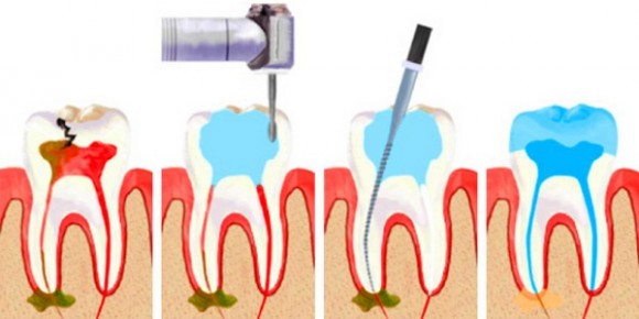
Treatment of purulent pulpitis
To treat purulent pulpitis, the neurovascular bundle is always removed, i.e. the causative tooth is depulped. The treatment protocol may include either a vital endodontic method or a non-vital method.
The vital method is performed in one visit under local anesthesia.
This procedure consists of the following steps:
First, the dentist administers anesthesia.
As soon as the anesthetic takes effect, the doctor begins mechanical cleansing of the carious cavity.
Medicinal irrigation of the cavity with an antiseptic solution.
Opening the pulp horn and extracting the neurovascular bundle with small endodontic instruments.
Expansion of the walls of the root canal with simultaneous mechanical cleaning.
Antiseptic treatment of the root canal.
If blood is detected from the canal, it is stopped with special solutions.
Dry the canal thoroughly with paper points.
Filling root canals and carious cavities.
Control x-ray to ensure high-quality obturation (closure).
Grinding and polishing of the filling.
Treatment of acute purulent pulpitis using the devital method is carried out in 2 visits. On the first visit, the tooth is numbed using the application method. The pulp horn is opened and a ball of devitalizing (pulp-killing) paste is placed on it. Previously, a paste containing arsenic was used for these purposes.
Modern dentistry has abandoned this drug, and paraformaldehyde plays a killing role. The doctor will place a little cotton wool on the paste, and a temporary filling on it.
The doctor recommends a second visit no earlier than after 10 days, so that the pulp is certainly mummified and endodontic manipulations do not cause pain. All other steps are completely identical to those described above, only local anesthesia is not needed in this case.


