The outer layer of enamel and the inner layer (5 - 15 µm) at the dentino-enamel border do not contain prisms (prismless enamel). These layers contain small crystals and larger lamellar crystals.
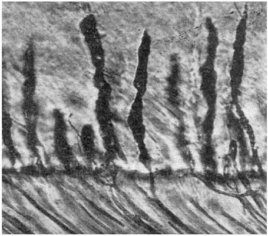
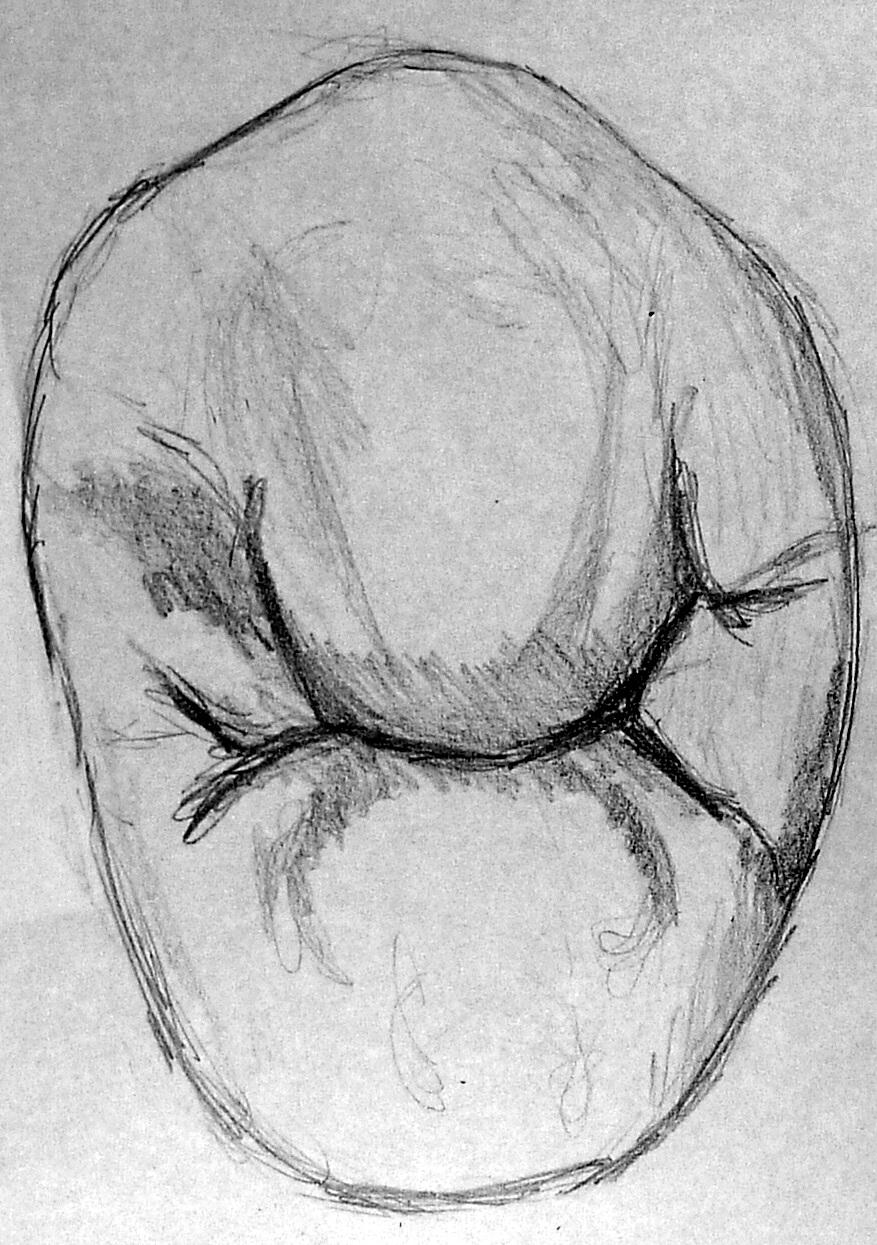
In enamel there are also enamel plates (lamellae) and bundles, which are areas of insufficiently mineralized interprismatic substance. The plates pass through the entire thickness of the enamel. The bundles are located mainly at the dentinoenamel border. These formations can serve as entry points for bacteria and starting points for the development of caries (Fig. 4.25).
Rice. 4.25. Enamel plates (1) and enamel bundles (2) in the enamel of a human molar. Transverse section of the tooth. (According to Falin L.I., 1963, M.)
The next structural element of enamel is enamel spindles- flask-shaped thickenings of odontoblast processes penetrating into the enamel through the dentin-enamel junction. The spindles are located between the enamel prisms and take part in the trophism of the enamel
The greatest thickness of enamel is in the area of the cusps (1.7 mm), the thinnest is in the area of the neck of the tooth (0.1 mm). The thickness of the enamel in the fissures of the chewing surface is 0.6-0.7 mm.
 Rice. 4.26. Enamel spindles are processes of odontoblasts penetrating through the enamel-dentin junction into the enamel (According to Falin L.I., 1963, M.)
Preparation of the enamel itself is painless, but processing it in the cervical area is often very sensitive due to the rapid penetration of the bur into the dentin (passage of the enamel-dentin junction). Due to its high mineralization, enamel is not cut with burs, but rather polished, so it is better to process it with grinding tools (diamond or carbide burs, carborundum stones). Along with high strength, enamel has significant fragility. This circumstance must be taken into account when forming cavities, i.e. in places of high mechanical load, overhanging and thinned edges of the enamel must be excised. For the same reasons, one should not undermine the enamel in the area of cusps and cutting edges of tooth crowns. The significant strength of enamel is associated with the crystalline structure of its prisms, which are based on crystals of hydroxyapatite (calcium phosphate). Enamel does not have regenerative properties, but it is characterized by the phenomenon of remineralization, i.e., ion exchange associated with the entry of calcium salts, phosphorus and trace elements into it from saliva. The phenomenon of enamel remineralization is used in clinical practice to increase the resistance of enamel to caries and other pathological processes. The latter is achieved by applying fluorine preparations, calcium salts and phosphorus to its surface. Despite its high mechanical strength, tooth enamel is easily destroyed by the action of some organic and inorganic acids. Enamel prisms in the area of chewing cusps and cutting edges lie parallel to the tooth axis, and on the lateral surfaces they gradually move to a plane perpendicular to the tooth axis. These features of the location of enamel prisms must be taken into account when preparing enamel.
(Fig. 1) has a prismatic crown, the buccal and lingual surfaces are convex. The vestibular surface is larger than the palatine surface and has a small vertical ridge. The contact surfaces are rectangular in shape, with the rear surface being more convex than the front. There are 2 tubercles on the chewing surface - buccal and palatal. The buccal area is much larger. Between the tubercles in the anteroposterior direction there are grooves (fissures), which end in small enamel ridges. On the chewing surface of the buccal tubercle, two slopes are distinguished, the anterior one being better expressed. The root is flattened, with deep longitudinal grooves on its lateral surfaces. The root often bifurcates into a buccal and a better defined palatal root. The signs are well expressed. However, the tooth often has the opposite sign of crown curvature, i.e. the posterior part of the buccal surface is more convex, the anterior part is more sloping.
(Fig. 2). It has a slightly smaller size. The crown is prismatic in shape. There are two tubercles on the chewing surface. Buccal and palatal. The buccal region is better developed. The tubercles are separated by a transverse groove running through the center of the chewing surface and separated from the edges of the crown by small enamel and ridges. The buccal surface of the crown is larger than the palatal surface. The palatine is more convex and has a longitudinal ridge. The anterior portion of the buccal surface of the crown is less convex compared to the posterior one (the opposite sign of crown curvature). The root is often single, cone-shaped, compressed in the anteroposterior direction, the lateral surfaces are wide, and have shallow longitudinal grooves. In 15% of cases, a bifurcation of the root is observed closer to the apex.
(Fig. 3). The vestibular surface of the crown is convex, longer than the lingual. On the vestibular surface there is a wide longitudinal ridge, heading towards the main tubercle of the chewing surface.
The chewing surface has two tubercles. The lingual cusp is always smaller than the buccal cusp. The buccal is larger, strongly inclined inward. They are separated by a small groove, which is located closer to the lingual tubercle. The tubercles are connected at the edges by a ridge, on the sides of which there are small depressions (pits). The root is single, straight, oval in shape, slightly flattened on the sides. There are shallow grooves on the front and back surfaces.
(Fig. 4) is larger than the first premolar.
 The vestibular surface is similar, but the lingual surface is slightly larger due to the well-developed lingual tubercle. The cusps are almost equally developed (the buccal ones are somewhat larger), separated by an enamel ridge, on the sides of which there are small depressions (pits). The ridge is separated from the tooth edges by a horseshoe-shaped fissure. An additional groove may extend from the fissure, which divides the lingual cusp into two smaller cusps, turning the tooth into a tricuspid one. The contact surfaces are convex and pass into the lingual surface without sharp boundaries. A longitudinal ridge runs along the lingual surface, ending at the lingual tubercle. The root is single, cone-shaped. Slightly flattened, its lateral surfaces are almost devoid of longitudinal grooves. The root sign is well expressed. Signs of angle and curvature are not clearly expressed.
Small molars (premolars)(dentes premolares). The small and large molars are lateral teeth. Their function is to chew and grind food, as a result of which these teeth are also called chewing teeth. Upper premolars often have two roots, and lower premolars often have one. The closure surface of the premolars is wide and, as a rule, has two tubercles - vestibular and lingual, which facilitate chewing food. Located between the canines and molars, the premolars have some characteristics of the teeth of neighboring classes.
Upper small molars (premolars). Typically, the upper premolars are larger than the lower ones. There are 1st and 2nd premolars.
1st upper premolar has a vestibular (buccal) crown surface, which is similar in shape to the canine crown (Fig. 1). The cutting edge of the crown bears the main tubercle in the middle, lower than that of the canines. The mesial and distal parts of the edge extend from the main tubercle at an obtuse angle. The contact surfaces move somewhat closer towards the neck. The enamel-cement border is arched and convexly directed towards the root. From the main tubercle of the cutting edge in the middle of the buccal surface of the tooth, a wide convex ridge, shaped like an elongated oval, extends to the neck. Narrow marginal ridges follow from the lateral corners of the crown to the neck, which can connect at the enamel border with middle roller. Two shallow grooves are noted between the marginal and median ridges. The mesial ridge is usually better developed than the distal one, and the mesial angle of the crown is well defined. Sometimes a small intermediate tubercle is found on the distal edge of the incisal edge.
Rice. 1. First upper premolar, right:
It is difficult to use the sign of the crown angle for the upper premolars, since almost equally often the rounded obtuse angle can be both the mesial and distal angle of the crown. The relationship of the ribs of the cutting edge is uncertain: in some cases the mesial edge is shorter and flatter than the distal one, in others, on the contrary, it is longer and steeper.
The mesial and distal contact surfaces of the crown form a small angle with the corresponding surfaces of the root. More often, the angle between the distal surfaces is greater than between the mesial ones, but quite often both of these angles are approximately the same, so the sign of root curvature for the upper premolars is not always reliable.
The root of the upper premolar is flattened in the mesiodistal direction. The root apices often deviate distally, but straight roots or even mesially deviated roots are found. Often a longitudinal groove is noticeable on the buccal surface of the root, and in rare cases the buccal root is split in two.
When examining the occlusal surface of the upper premolar, two things are clearly visible: masticatory cusps- buccal, larger, and lingual, somewhat smaller. Sometimes both tubercles are developed approximately equally. Between them lies quite deep intertubercular groove (sulcus intertubercularis), which does not reach the lateral edges of the crown. Along the edges of the chewing surface of the crown there are edge scallops- mesial and distal. Each consists of two parts: the vestibular, arising from the vestibular tubercle of the occlusal surface, and the lingual, arising from the lingual tubercle. Towards the middle of the lateral edges of the crown, the height of the ridges decreases, but they still limit the intertubercular groove. The latter can cut through the lateral ridges, most often the mesial one. In addition, it usually bifurcates at the mesial end, giving off a small groove vestibularly. In such cases, an intermediate mesial tubercle is formed between the branches of the groove, which may indicate that the tooth belongs to one or another half of the dental arch. The division of the intertubercular groove at its distal end and the formation of an intermediate distal tubercle are extremely rare.
The inclination of the buccal and lingual cusps is expressed differently and can be steeper or gentler. The marginal ridges are also unequally expressed, and the ridges adjacent to the buccal tubercle of the occlusal surface are usually larger than those going to the lingual tubercle. Meet extra scallops, usually on the distal side. The depth of the intertubercular groove is associated with the development of the ridges. The furrow can be very deep, medium or shallow. With steep inclinations of the buccal and lingual cusps along the intertubercular groove, which in such cases is wide, there may be additional grooves and additional central tubercles- mesial and distal. An important feature of the upper premolars is mesial shift of the lingual tubercle.
The lingual surface of the upper premolars is usually smooth. The enamel-cement border on the buccal and lingual surfaces runs in an arcuate convex manner towards the root.
The approximal surfaces of the crown are more or less convex. In the middle of both the mesial and distal surfaces there can be a longitudinal groove, corresponding to the intertubercular groove of the occlusal surface, which divides the crown into two parts. Sometimes ridges spread from the tubercles along the lateral surface of the crown. Most common lingual ridge on the distal surface. The enamel-cement border on the lateral surfaces comes in different shapes. With one root, the border is located in an arched manner with a convexity towards the chewing surface, and the greatest height of the arc falls on the buccal tubercle. Less often the border goes straight. With two roots, the enamel border has two bends, open to the root. The higher curve corresponds to the buccal tubercle. Between the bends, corresponding to the interradicular groove, there is a protrusion of enamel facing the apex of the root, which can sometimes spread downward in the form of a narrow strip, forming an interradicular enamel streak. The contour of the buccal surface of the crown of the upper premolars can be uniformly convex or inclined in the lingual direction. In the cervical area, the contour of the crown from both the buccal and lingual surfaces can rise. The contour of the lingual surface has similar features. Depending on the severity of the inclination of the surface, the crown of the upper premolars may be more or less open. In the lateral norm, the relationship between the buccal and lingual cusps is clearly visible:
a) the buccal tubercle is significantly higher in height than the lingual one;
b) the lingual tubercle is slightly smaller than the buccal tubercle;
c) both tubercles are the same size.
Upper premolars can have 1, 2 and extremely rarely 3 roots. A single root tapers wedge-shaped towards the apex, its lateral contours are convex or almost straight; There are longitudinal tubercles in the middle of both root surfaces. The root apex may be deviated lingually or mesially. The differentiation of the root system can be weak - grooves on the mesial and distal surfaces of the root; medium - partial splitting of the root into two; strong - the formation of two roots; extremely strong - the formation of three roots.
The cavity of the crown of the upper premolars is quite large, more or less cylindrical in shape, and has two protrusions corresponding to the masticatory tubercles. The buccal protrusion is usually longer than the lingual. At the base of the crown, the root canals also enter the cavity. The palatal root canal is usually wider than the others. With one root, its canal is compressed in the mesiodistal direction.
1st upper premolar, as a rule, has two roots - buccal and lingual. The height of the crown along the buccal surface is 7.5-9.0 mm, along the lingual surface - from 6 to 8 mm, the width of the crown in the widest part of the buccal surface is 6.5-7.0 mm, the mesiodistal size of the crown is 4.8-5 .5 mm, buccolingual - from 8.5 to 9.5 mm; root length: palatal - 12.5-15.5 mm, buccal - 12.5-14.0 mm.
2nd upper premolar very similar to the 1st (Fig. 2), the relief of the crown is smoothed, its vestibular surface is often oval. The cutting edge of the crown has rounded corners, the chewing tubercles on the contact surface are more or less equal in height. Edge scallops and ramifications intertubercular groove poorly developed, additional central tubercles on the chewing surface they are very rare. The 2nd upper premolar often (90%) has one root and one root canal, less often (10%) 2-3 roots.
Rice. 2. Second upper premolar, right:
a — vestibular surface; b — mesial surface; c - lingual surface; d — vestibular-lingual section; d — mesiodistal section; c - cutting edge; 1, 2, 3 - shape of cross sections at the level of the crown, middle and upper third of the root, respectively
The height of the crown on the buccal surface is 7.5-8.5 mm, on the lingual surface - from 6.5 to 7.5 mm, the width of the crown is 6-7 mm, the mesiodistal size is 4.5-5.5 mm, buccal lingual - from 8 to 9.5 mm; root length - 13.0-16.5 mm.
The upper premolars are located in the upper dental arch in a uniformly rounded area. The 2nd upper premolar is more variable, and the 1st is a relatively stable tooth. Maybe congenital absence 2nd upper premolar, and there is also a 3rd premolar, which can erupt both within the dentition and outside it.
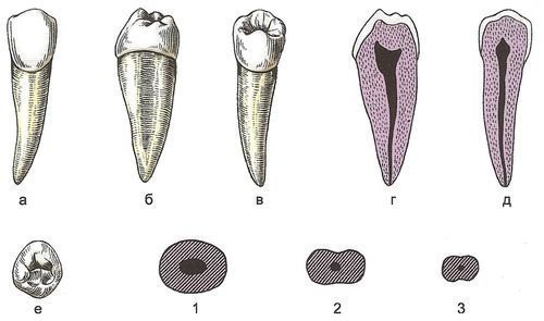
Lower small molars (premolars). The crowns of the lower premolars are smaller than the crowns of the upper premolars, but the root length is longer. Usually there is one root. The 1st and 2nd lower molars differ in structure more than the upper ones.
1st lower premolar the shape of the crown is very similar to the canine (Fig. 1). On the vestibular (buccal) surface on the cutting edge there is a main cusp, which is usually lower than that of the canines. The angle between the sections of the cutting edge forming the tubercle is obtuse. There are 1st premolars with a high cusp, very similar to canines. The mesial rib is usually shorter and more shallow than the distal rib. The distal corner of the crown is rounded. Sometimes angular tubercles are noticeable at the corners of the crown; the cutting edge in these cases appears to be three-tubercle. A wide longitudinal ridge runs along the occlusal surface of the crown from the main tubercle towards the neck, which gradually decreases and disappears in the middle third of the crown. From the corners the crowns are small and short corner scallops. The lateral edges move somewhat closer towards the base of the crown.
Rice. 1. First lower premolar, right:
a — vestibular surface; b — mesial surface; c - lingual surface; d — vestibular-lingual section; d — mesiodistal section; e - cutting edge; 1, 2, 3 - shape of cross sections at the level of the crown, middle and upper third of the root, respectively
The vestibular surface of the crown is convex in the horizontal plane, and its curvature is increased in the mesial half of the crown.
The occlusal surface of the lower premolars may have a different structure due to the variability of the structure lingual dental tubercle. With a canine-shaped premolar, the lingual tubercle is poorly developed and difficult to distinguish from median scallop, running from the main tubercle of the cutting edge to the lingual tubercle. There are two pits on either side of the scallop. In other cases, the lingual cusp is large, and the occlusal surface takes on a two-cusp shape characteristic of premolars. In this case, a deep groove passes between the vestibular and lingual tubercles, cutting the median vestibulolingual ridge. Rarely, the lingual cusp is divided into two, and then the occlusal surface of the premolar becomes tricuspid. Each of the two lingual cusps passes into the marginal ridges of the occlusal surface.
When viewing the tooth in lateral projection, the vestibular contour of the crown is almost straight and strongly deviates in the lingual direction. The contour of the lingual surface is also straight, its occlusal edge hangs over the base of the crown. A noticeable transverse coronal groove.
The lingual surface of the crown of the 1st premolar is convex, its edges evenly approach the neck. In the middle of the cutting edge, the lingual tubercle rises.
The root is often single, sometimes double, but complete splitting of the root is rare. A single root is compressed in the mesiodistal direction, its buccal surface is wider than the lingual, and sometimes bears a longitudinal groove. The root is deviated distally. The middle sections of both sides of the root are not always pronounced. The contours of the root are straight or slightly convex. The enamel-cementum border runs in an arcuate manner, and the border at the vestibular surface extends onto the root more than that of the lingual surface.
With two roots, the mesial one is shifted in the buccal direction, and the distal one is shifted in the lingual direction. Both roots are flattened and sometimes have longitudinal grooves.
The cavity of the crown of the lower premolars is rounded, has two horns corresponding to the tubercles of the occlusal surface. The root canal is wide and sometimes bifurcates.
The height of the crown of the 1st lower premolar on the buccal surface is 7.5-11.0 mm, on the lingual surface - from 5 to 6 mm, crown width - 6-8 mm, buccolingual neck diameter - 8.2-8.6 mm, mesiodistal - 5.4-5.8 mm; root length - 13.0-16.5 mm.
2nd lower premolar has a hemispherical crown. The buccal surface is more even (Fig. 2). Median ridge coming from main tubercle cutting edge, wide and relatively flat.
Rice. 2. Second lower premolar, right:
a — vestibular surface; b — mesial surface; c - lingual surface; d — vestibular-lingual section; d — mesiodistal section; e - cutting edge; 1, 2, 3 - shape of cross sections at the level of the crown, middle and upper third of the root, respectively
The main vestibular tubercle is lower than that of the 1st premolar; the cutting edge ribs that form it converge at an obtuse angle, with the mesial rib being shorter than the distal one. The distal corner of the cutting edge is rounded, sometimes bearing a small intermediate tubercle. The edges of the buccal surface of the crown approach slightly at the neck. The enamel-cementum border is arched and open to the cutting edge.
The occlusal surface is often bicuspid. The lingual tubercle is very well developed and only slightly lower than the buccal tubercle. There are teeth with tubercles of equal size. The occlusal surface can be tricuspid (division of the lingual cusp into two), quadricuspal (division of the distal angular cusp, shift of the main vestibular cusp in the mesial direction and separation of the intermediate cusp on the distal edge of the buccal cutting edge). Between the buccal and lingual elevations of the occlusal surface there is a deep transverse groove, which has terminal branches.
The approximal surfaces of the crown (mesial and distal) are shaped like a hemisphere. In the 2nd premolar, unlike the 1st, the contours of both the buccal and lingual surfaces of the crown have the form of arcs of large radius or straight lines, with bevels to the occlusal surface. The buccal and lingual elevations are clearly visible and are almost the same height. The enamel border on the buccal surface lies lower than on the lingual surface, and on the contact surface it is a gentle arc open to the apex of the tooth.
The lingual surface of the crown is smooth and convex.
The root of the tooth is usually single. It is longer than that of the 1st premolar, its surfaces are smooth and convex. Longitudinal grooves on the lateral surfaces are rare, the apex is deviated distally.
The cavity of the crown of this tooth is cylindrical in shape, its lingual horn is larger than that of the 1st premolar. The root canal is wide and long.
The height of the crown on the buccal surface is 7-9 mm, on the lingual surface - from 6.5 to 9 mm, mesiodistal size - from 4.5 to 6.5 mm; root length - 14.0-17.5 mm.
The 2nd premolar is variable. All transitional forms of the occlusal surface of this premolar to the shape of the surface of the large molars are observed. Possible congenital absence 2nd premolar and crowding. Tremas most often occur between the canine and the 1st premolar, very rarely between premolars.
The upper and lower premolars are in contact with each other in such a way that each lower premolar falls on two adjacent upper ones. Thus, the 1st lower premolar closes with the distal half of the crown of the upper canine and with the mesial half of the 1st upper premolar; The 2nd lower premolar occludes with the distal half of the 1st upper premolar and with the mesial half of the 2nd upper.
Human anatomy S.S. Mikhailov, A.V. Chukbar, A.G. Tsybulkin
Lesson 13
Particular anatomy of permanent dentition.
Premolars of the upper jaw.
Learning objective: To teach students the features of the anatomical structure of the premolars of the upper jaw. Learn to find the distinctive features of the first and second premolars, the teeth belonging to the right and left sides.
Lesson duration: 2 hours
Location of the lesson: clinical base
Practical lesson plan:
|
Lesson stages |
Equipment |
Visual aids | |
|
1. Checking the presence of students | |||
|
2. Teacher briefing | |||
|
3. Test control of initial knowledge |
Test tasks | ||
|
4. Oral control of initial knowledge |
Control questions | ||
|
5. Analysis of the practical lesson topic |
Practical guides, teaching aids | ||
|
6. Working with manual skills |
Material for sculpting artificial teeth and tools |
Slides, pictures, presentations | |
|
7. Monitoring the assimilation of material |
Questions to monitor the results of mastering the material | ||
|
8. Assignment for the next lesson |
Questions studied previously and necessary for the lesson:
Differences in the anatomical structure of the canines of the upper and lower jaw
Determining whether fangs belong to the right and left sides
Functional significance of canines
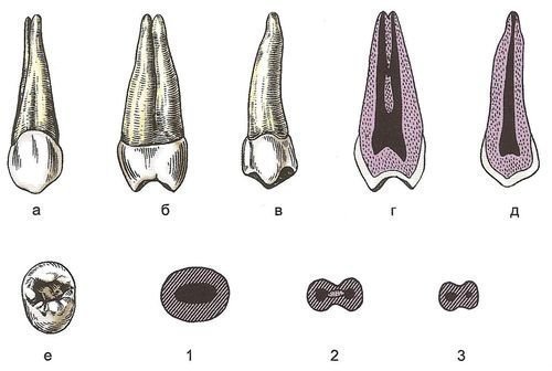
Maxillary first premolar
It has the shape of a crown - rounded rectangular.
There are three ridges on the vestibular surface: longitudinal, medial, distal, with two depressions: medial, distal.
From the oral surface I also distinguish three ridges: longitudinal, medial, distal.
On the lateral surfaces there are two tearing tubercles: vestibular, palatine, two ridges: medial/distal palatine, medial/distal vestibular, intertubercular groove.
There are six ridges on the occlusal surface: longitudinal vestibular, longitudinal palatine, medial vestibular, distal vestibular, medial palatine, distal palatine, additional medial tubercle, intertubercular groove with 2 branches (medial and distal).
The cavity of the crown is large, narrowed in the medio-distal direction, and has two protrusions corresponding to the masticatory cusps.
The root is flattened, with deep longitudinal grooves on the lateral surfaces. The root often bifurcates into a buccal and palatal root.
The signs are well expressed.
Crown height on the buccal surface 7.5 - 9 mm
Crown height on the lingual surface 6 - 8 mm
Width 6.5 - 7 mm
Medial-distal diameter of the tooth neck 4.8 - 5.5 mm
Buccal-lingual diameter of the tooth neck 8.5 - 9.5 mm
 Root length 12 - 16 mm
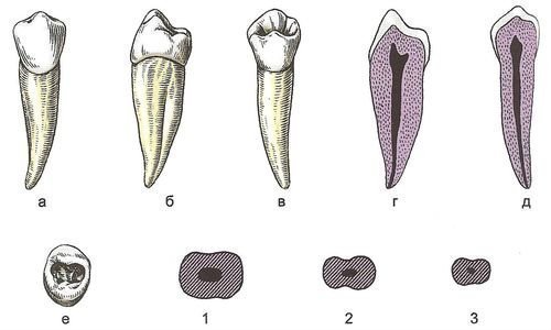
Maxillary second premolar
Slightly different from the first one, but somewhat smaller in size.
On the vestibular surface there are four ridges: longitudinal, medial, distal, additional distal, forming three depressions: medial, distal, distolateral, tearing tubercle.
Three ridges are distinguished from the oral surface: longitudinal, medial, distal, and tearing tubercle.
From the lateral surfaces there are four ridges: longitudinal vestibular, longitudinal palatine, medial distal vestibular, medial distal palatine, separated by two branches of the intertubercular fissure: palatine, vestibular.
There are six ridges on the chewing surface: longitudinal vestibular, longitudinal palatine, medial vestibular, distal vestibular, longitudinal medial, longitudinal distal, separated by an intertubercular fissure.
The root is often single, cone-shaped, compressed in the anteroposterior direction, the lateral surfaces are wide, and have shallow longitudinal grooves. In 15% of cases, a bifurcation of the root is observed closer to the apex.
Crown height on the buccal surface 7.5 - 8.5 mm
Crown height on the lingual surface 6.5 - 7.5 mm
Width 6 - 7 mm
Medial-distal diameter of the tooth neck 4.5 - 5.5 mm
Buccal-lingual diameter of the tooth neck 8 - 9.5 mm
 Root length 12.5 - 16.5 mm
Questions to control the assimilation of the material:
1.Name the distinctive features of the first and second premolar of the upper jaw;
2.What surfaces are distinguished in premolars;
3.What shape do the premolars have on the lateral surfaces?
4.what shape does the tooth cavity have?
5.What fissures are distinguished on the first and second premolars of the upper jaw.
Literature
Phantom course of therapeutic dentistry. Atlas. Yu.M. Maksimovsky, textbook. Benefit. –M. “Medicine”, 2005. – 328 p.
Propaedeutic dentistry.
M.M. Pozharitskaya, T.G. Simakova, M., “Medicine”, 2004. – 304 p.
Orthopedic Dentistry Ed. V.N. Trezubov.-7th ed., revised.
And additional – St. Petersburg: Foliant, 2006. – 592 p.
Surgical dentistry Robustova
Phantom course of therapeutic dentistry. A.I. Nikolaev, L.M. Tsepov “Medpress-inform” M. 2009., 430 p.
Propaedeutic dentistry. Textbook. Edited by Professor E.A. Bazikyan.
M. "GEOTAR-Media", 2010.
“Anatomy of human teeth” I.V. Gaivoronsky, T.B. Petrova;, or premolars, denies premolares, - include 8 teeth in their group; four on each jaw, two on the right side and two on the left. Small molars are divided into first and second premolars. The first is called proximal, and the second is called distal. The upper molars (premolars) have an oval-shaped crown and two roots. The second upper premolar in 80% of cases has one root (S. S. Mikhailov, 1973). The root is compressed in the mediodistal direction and has a longitudinal groove. There are two chewing tubercles on the chewing surface: parietal and lingual.
The lower molars (premolars) have a crown and a longer root.
Morphological characteristics of small molars (premolars)
First upper molar(first upper premolar) is characterized by an irregular crown of a prismatic shape, in which the buccal surface is always wider than the lingual surface, the diameter of the crown is larger in the buccolingual direction. The cheek surface is convex, with clearly defined signs of crown curvature, which in these teeth can often be reversed, that is, the back part of the surface is more convex, and the front part is flatter. The buccal surface, forming rounded corners, becomes contact. They are rectangular in shape and swollen, with the distal surface being more swollen. These surfaces (distal and medial), without forming a forged surface, smoothly transform into a convex lingual surface. The chewing surface is formed by two tubercles, of which the buccal one is slightly larger. Between the tubercles there is a fissure, which is limited at the edges by small transverse grooves, as a result of which rolls are formed at the edges of the chewing surface. The first upper premolar has a somewhat peculiar root structure, which is completely or partially divided into two roots - buccal and lingual. The limit of root division is different: it is mainly localized at the apex of the root, but can also be localized in the middle of the root or closer to the cervical part of the tooth. The higher the limit of root splitting is localized, the more the tubercles of the chewing surface converge.The tooth cavity is located in the cervical part of the crown and root, compressed in the approximal-distal direction; has the shape of a slit. It is formed by five walls; the bottom of the chamber is a convex bifurcation of the roots, through which the cavity continues in two root canals. In frontal and sagittal sections, the tooth cavity vaguely repeats the shape of the crown in a reduced size. In a cross-section directly under the arch, the tooth cavity follows the shape of the chewing surface and has two horns corresponding to the tubercles of the chewing surface in the bucco-palatal direction.
In the first upper small molars (premolars) all the anatomical features are clearly expressed, by which the teeth of the right or left half of the jaw are determined.
Second upper molar(second upper premolar) is similar to the first upper molar, but its crown is slightly smaller than that of the first premolar. Both mounds of the chewing surface are equally developed. The differences in characteristics are clear. The root is one, cone-shaped, its lateral surfaces are compressed from the sides. Sometimes there is a partial bifurcation of the root at the apex.
The tooth cavity (pulse chamber) of the second premolar is similar to the cavity of the first premolars and, gradually narrowing, often turns into one root canal. However, in approximately 40% of cases, two canals are observed, which often merge at the apex into one.
First lower molar(first lower premolar) has a round crown, somewhat narrowed in the buccal direction. There are two tubercles on the chewing surface: a large one, slightly inclined into the oral cavity (buccal), and a small one (lingual). The tubercles at the front and back surfaces are connected by enamel rollers. The same ridge sometimes runs from the middle of the buccal tubercle to the lingual tubercle, and then two pits are formed on the sides. It happens that the chewing surface in the anteroposterior direction has a groove located closer to the lingual edge of the crown, then the lingual tubercle is always smaller.
The buccal surface is similar in shape to the surface of the lower canine; its chewing edge in the middle has a tooth corresponding to the top of the buccal tubercle and divides it into medial and distal segments. The distal segment is longer than the medial one. The lingual surface is smaller and lower than the buccal surface and smoothly passes into convex contact surfaces. Due to its anatomical and topographical features, the first lower premolar is similar to the lower canine. It has a single rounded root, somewhat elongated in the buccal-lingual direction, with not very pronounced grooves on the anterior and posterior surfaces. Often the crown and root are located at an obtuse angle to each other, with the crown inclined towards the tongue. All signs of differences in the teeth of the first lower premolar are well expressed.
The dental cavity in sagittal and frontal sections somewhat repeats the shape of the teeth in reduced sizes. In a cross section directly under the arch, it has the shape of an oval, which, gradually narrowing, transforms into a well-defined root canal.
Second lower molar(second lower premolar) - has a rounded crown, due to the greater development of the lingual tubercle, the crown of the first premolars is larger. The chewing surface of the second premolar has two equally developed tubercles; on the sides between the tubercles there are enamel ridges and a deep groove; often an additional groove extends from it, dividing the lingual tubercle into two tubercles, and then the tooth becomes tricuspid. The buccal surface does not differ from the same surface of the first premolar, but the contact surfaces are somewhat large, convex and gradually transform into the lingual surface. The lingual surface, due to the well-developed lingual mound, is also larger than the lingual surface of the first premolar. The root of the second lower premolar is cone-shaped and slightly longer than the root of the first lower premolar. The angle sign is weakly expressed, the root sign is better expressed.
The tooth cavity is located in the cervical part of the tooth crown and at the beginning of the root; gradually narrowing, it transforms into one, well-defined channel. On a cross-section, directly under the chewing surface, the tooth cavity repeats the shape of the chewing surface, and at the level of the neck it has an oval shape.


