One of the reasons for the body’s own lymphocytes to attack the body’s cells may be the similar structure of the body’s cells with the antigens of a bacterium or virus, i.e. the lymphocyte “confuses” its own cells with the antigens of infectious agents.
As a rule, the tendency to autoimmune pathology is genetic. Predisposing factors may include UV irradiation, infections, uncontrolled and unjustified use of immunostimulating agents, exposure to any chemicals.
Nature autoimmune diseases in cats has not yet been studied enough. With pemphigus, dysfunction immune system the animal leads to an attack of its own epidermal cells. The destruction of skin cells and the release of their contents is clinically manifested by the formation of blisters.
One of the reasons for the body’s own lymphocytes to attack the body’s cells may be the similar structure of the body’s cells with the antigens of a bacterium or virus, i.e. the lymphocyte “confuses” its own cells with the antigens of infectious agents.
The second reason may be a violation of the screening of autoreactive lymphocytes at the stage of their maturation. If a lymphocyte at the maturation stage is not able to distinguish the cells of the host body from foreign antigens, then such a lymphocyte must be destroyed. Sometimes the destruction mechanisms are disrupted.
Autoimmune antibodies: the body produces antibodies that attack healthy tissues and cells as if they were pathogenic.
Prolonged exposure to the sun.
Some breeds may have a hereditary predisposition.
Types of pemphigus
There are 4 types of pemphigus, affecting dogs: pemphigus foliaceus, pemphigus erythematous, pemphigus vulgaris and pemphigus vegetans.
In pemphigus foliaceus, autoantibodies are found in the outermost layers of the epidermis and blisters begin to form on the healthy skin. Erythematous pemphigus occurs in almost the same way as foliaceous pemphigus, but is less painful.
Pemphigus vulgaris is characterized by the formation of deeper ulcers, as antibodies accumulate in the lower layers of the epidermis. As for pemphigus vegetans, it affects only dogs and is considered the rarest variety.
Pemphigus vegetans resembles pemphigus vulgaris, but is much milder with the formation of less painful ulcers.
Clinical signs
Since exfoliative pemphigus is most common in cats, we will first look at the symptoms of this type of disease:
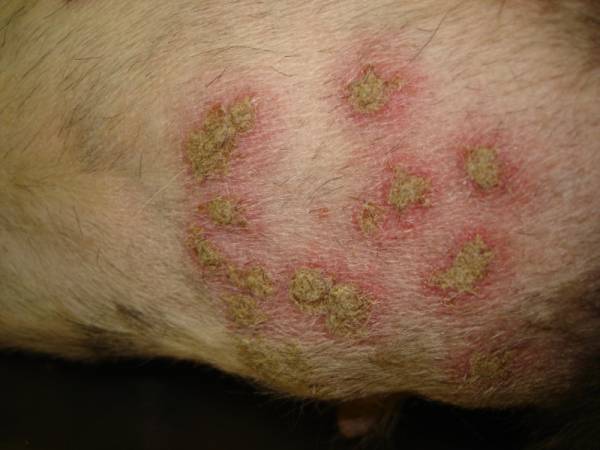
- Generalized rashes of pustules (pictured), multiple crusts, small ulcers, redness and itching of the skin, including the head, ears and groin area are most often affected.
- In other cases, large papules filled with cloudy liquid are observed.
- Large cysts often form in the thickness of the skin.
- In severe cases, the gums are also involved in the process, resulting in problems with teeth (even tooth loss).
- Similarly, the nail beds are involved in the process, the animal’s claws begin to wobble and sometimes fall out. The process is very painful and causes great suffering to the animal.
- Swollen lymph nodes; when palpated, the cat clearly shows signs of displeasure. The animal becomes apathetic, with increasing fever and lameness (if claws are involved). Note that all these signs are characteristic only for severe course process.
- Secondary bacterial infection is possible due to the contamination of opened papules and ulcers with pyogenic microflora.
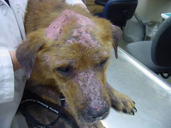
This is the name of an autoimmune skin disease that is most often detected in young and middle-aged dogs. The main symptom of the disease is multiple pustules and crusts covering the surface of the affected areas of the skin. Maceration and severe pain of the latter are also characteristic. The disease can begin on the face and ears, but cases of involvement of the groin and axillary areas are also common. The diseased skin thickens significantly and may crack, causing severe pain. Fortunately, internal organs the disease does not affect.
What causes pemphigus in general, what are the triggering mechanisms of this pathology? As we have already mentioned, the disease is autoimmune, that is, it occurs due to the fact that the immune system fails and begins to attack the body itself. The consequences are very serious, since there is not even a full-fledged treatment for such cases: doctors simply extinguish the main symptoms and struggle with the consequences. In the case of pemphigus, the only consolation is that no internal organs or systems pathological process are not affected. If you compare it with the same systemic lupus, then pemphigus is tolerated by animals much more easily.
What causes the immune system to attack the body itself? Unfortunately, we don't know for sure. There are probably many predisposing factors, but which one plays the role of a trigger in each specific case? Most likely, infectious diseases, genetic pathologies, and some medications are very dangerous in this regard. Today, many veterinarians and breeders believe that pemphigus can be inherited. That is why those animals that are sick with it should under no circumstances be allowed into breeding, even if their breeding value is very high.
Read also: Cerebral edema in dogs: causes, types and symptoms
Symptoms
Symptoms of pemphigus in a dog largely depend on the type of disease. There are four in total:
- Common (vulgaris).
- Erythematous.
- Leaf-shaped.
- Vegetative. The latter type affects dogs exclusively (but extremely rarely).
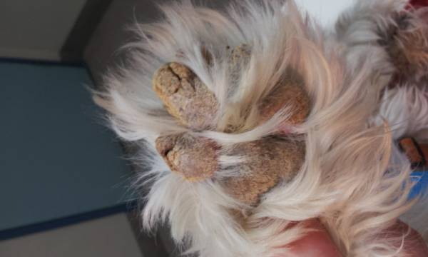
So, by the presence of what signs can one judge the presence of the disease? Numerous ulcers, pustules and vesicles filled with cloudy contents appear on the surface of the skin. The paw pads are also affected and become prone to cracking and inflammation (the photo shows just such a case).
If the disease is severe, it is accompanied by inflammation and swelling lymph nodes, the animal is depressed, cases of intermittent fever cannot be excluded. In cases where the paw pads are affected, the animal limps severely, trying not to move at all unless absolutely necessary. All this is accompanied severe pain and itching.
All types of pemphigus are also dangerous because the sick dog’s body becomes especially susceptible to all secondary bacterial infections. Considering general weakness animal, one should not be surprised at the increased likelihood of sepsis.

Pemphigus vulgaris, that is, ordinary, is especially difficult. This type of disease is characterized by the formation of deep and very painful ulcers, “massive” ulcers and pustules. Since they often occur on the mucous membrane oral cavity, the animal cannot drink and eat normally. Almost always, it is pemphigus vulgaris that is accompanied by the development of secondary bacterial infection and fever, there is a high probability of sepsis. Pemphigus vegetans occurs most easily.
Read also: Joint dysplasia in dogs: details about the pathology
Diagnosis and treatment
The only one reliable way diagnosis - biopsy of the affected skin area followed by microscopic examination. In some cases, it is possible to cut off a suitable piece “live”, but this should not be done. The reason is that to obtain the data necessary for staging, it is necessary to take the skin at the border of diseased and healthy tissue. So the veterinarian has to use drugs for local or general anesthesia.
How is pemphigus treated in dogs? Alas, but the only one more or less effective method consists of prescribing drugs that suppress the immune system. Prednisone most often plays this role. It works quite quickly and effectively, but the problem is serious side effects. The first two weeks the medicine is administered into loading doses, after which for a month (or a month and a half) the dosage is reduced to the minimum possible.
The goal of therapy is to use the lowest possible dose of medication, balancing on the edge where the manifestations of the disease are still visible, but have no effect. negative influence on the quality of life of the pet. A small amount of scabs on a dog’s skin is better than a damaged liver and kidneys. But this can really happen if you overdo it with prednisolone! To avoid this, blood tests are needed to help monitor the condition of internal organs.
Other notes
However, your dog may be prescribed other drugs that suppress the immune system. Often their effect is weaker than that of prednisolone, but the dosage of the latter when taken can be significantly reduced. This is done to reduce the likelihood of side effects.
Diseases of the immune system of dogs
IMMUNODEFICIENCY CONDITIONS
Definition. Immunodeficiency states are disorders of the immune system, manifested by a deficiency of humoral and cellular immunity factors.
Causes, clinical signs and development of the disease.
Immunodeficiencies can be congenital or acquired. Congenital immunodeficiencies are associated with underdevelopment of one or another immunocompetent unit: T-lymphocytes, B-lymphocytes and stem cells. Clinically, these disorders are manifested by a decrease (less often, an increase) in the concentration of immunoglobulins in the blood, thymic hypoplasia, lymphopenia, thrombocytopenia, anemia, granulomatosis, retardation of growth and development. Puppies with congenital immunodeficiency usually die in suckling period or in the first half of life. In addition, there may be congenital disorders phagocytic system (impaired lysis of bacteria by phagocytes) and complement system.
The causes of acquired immunodeficiencies are radiation injuries, hemoblastosis (see), anemia (see), hyperadrenocorticism (see), severe infectious and invasive diseases, chronic intoxication, the use of cytostatics, glucocorticoids, non-steroidal analgesics; nutritional dystrophy; hypovitaminosis; artificial feeding. The puppies' body resistance to infections decreases somewhat approximately 3 weeks after weaning from the mother, due to the loss of colostral immunity.
The clinical picture of acquired immunodeficiencies consists of signs of the underlying disease and the immunodeficiency itself. In any case, such conditions are characterized by an increased incidence of infectious and invasive diseases, including those caused by opportunistic and saprophytic microflora, as well as increased risk the occurrence of tumors. Usually acquired immunodeficiency is a reversible condition, with the exception of hematological malignancies, malignant tumors anterior lobe of the pituitary gland and adrenal glands.
Diagnostics. In addition to reduced body resistance to infectious and invasive agents, immunodeficiencies are characterized by changes in clinical and immunological blood parameters: leukopenia (with hemoblastoses, leukocytosis and the appearance of blast cells are possible), hypoimmunoglobulinemia, a decrease in the concentration of T- and B-lymphocytes, a decrease in the phagocytic activity of peripheral blood neutrophils.
Treatment. For congenital immunodeficiencies, treatment is futile. Therapy for acquired immunodeficiencies consists of the following components: - elimination causative factor And pathogenetic therapy; - use of immunostimulants; - limiting contacts with possible sources of infection and infestation; - use of chemotherapeutic agents, vitamins, probiotics.
The following are used as immunostimulants:
Kinoron (contains leukocyte interferon and cytokines) 1-2 ampoules subcutaneously or intramuscularly 1 time every 2 days;
Levamisole orally at a dose of 0.5-2 mg/kg twice a week for 1-2 months;
· methyluracil orally at a dose of 10 mg/kg 2-3 times a day for 1 month;
· sodium nucleinate orally at a dose of 10 mg/kg 3 times a day for 1-2 weeks;
Dibazole orally at a dose of 0.1-0.5 mg/kg once a day for 1 month;
· prodigiosan intramuscularly in a dose of 0.5-1 ml of 0.005% solution once a week for 1-2 months;
· tactivin subcutaneously at a dose of 2 mcg/kg once a day for 1-2 weeks;
· timoptin subcutaneously at a dose of 2-5 mcg/kg once a day for 1-2 weeks;
· Thymalin intramuscularly at a dose of 0.5 mg/kg once a day for 7-10 days.
For prevention infectious complications antibiotics are used, for example bicillin-3 and 5.
Prevention of congenital immunodeficiencies involves culling producers whose offspring are diagnosed with them. Acquired immunodeficiencies are prevented by ensuring adequate feeding and maintenance of dogs, timely vaccinations and deworming.
AUTOIMMUNE DISEASES
DERMATOMYOSITIS
Definition. Dermatomyositis is an autoimmune disease characterized by inflammatory lesions mainly skin and muscles. Mostly shepherd dogs are affected.
Causes and development of the disease. The disease is usually preceded by a viral infection. It is believed that autoantibodies and circulating immune complexes affect the connective tissue of the skin, blood vessels, muscles, joints. As a result, alternatives arise - inflammatory processes, growing connective tissue, calcium salts are deposited, which leads to fibrosis and calcification various organs and fabrics.
Clinical signs. Dermatomyositis is characterized by symptoms of myositis, arthralgia, contractures, and various skin lesions.
The appearance of dense, round plaques on the skin, closely fused with the underlying tissues, and foci of hyperkeratosis is possible. The dog is lethargic, reluctant to move, and has a reduced appetite. Conjunctivitis, bronchopneumonia, and chronic diarrhea are often noted.
Diagnosis is based on clinical signs and the course of the disease.
Treatment. Glucocorticoids and non-steroidal anti-inflammatory drugs are prescribed (see "Lupus erythematosus").
Additionally, vitamins and anabolic steroids are used, for example retabolil.
Limit feeding the dog food containing increased amount calcium (bones, cottage cheese).
Prevention consists of timely vaccination and deworming of animals, which reduces the risk of autoimmune diseases.
RHEUMATOID ARTHRITIS
Definition. Rheumatoid arthritis is an autoimmune disease characterized by chronic inflammation limb joints.
Causes and development of the disease. Due to the damaging effects of circulating immune complexes, inflammation of the synovial membranes of the joints develops, cartilage is damaged and articular surfaces epiphyses, which can lead to persistent dysfunction of the musculoskeletal system. Rheumatoid arthritis is usually preceded by bacterial infections.
Clinical signs. Characteristic signs of symmetrical arthritis of small joints, especially fingers. They become swollen and painful. The dog limps, moves reluctantly, but, “running away,” stops limping. Sometimes other organs are affected and the course of the disease resembles lupus erythematosus (see).
Diagnostics based on clinical signs, medical history, and course of the disease. Additional information can give a blood test: characteristic increased ESR and availability rheumatoid factor(in 40-75% of affected dogs).
Treatment. The main treatment method is anti-inflammatory therapy. The dog is treated with ibuprofen, indomethacin, piroxicam, acetylsalicylic acid, analgin. If there is no improvement, proceed to the use of glucocorticoids (prednisolone, dexamethasone, triamcinolone). If an infection is suspected (conjunctivitis, fever), antibiotics are prescribed. At the same time, multivitamins can be administered, and Rumalon can be administered to stimulate the regeneration of articular cartilage. The dog is limited in physical activity.
Prevention. Necessary timely treatment acute and chronic infections.
MYASTHENIAS AUTOIMMUNE
Definition. Autoimmune myasthenia is a disease caused by a violation of muscle innervation due to the destruction of acetylcholine receptors by autoantibodies.
Causes and development of the disease. The immediate cause of the disease is a decrease in the number of acetylcholine receptors on the postsynaptic membrane of neuromuscular synapses due to their damage by autoantibodies. As a result, muscle tone decreases and the functions of various organs are disrupted.
Clinical signs. Dogs aged 3 to 10 years are most often affected. After a short physical activity animals get tired quickly, they notice muscle tremors. Data laboratory research within normal limits.
Diagnostics. After the use of anticholinesterase drugs, a temporary improvement occurs, which is a pathognomonic sign.
Treatment. Assign anticholinesterase drugs(prozerin, oxazil), as well as glucocorticoids.
Prevention not developed.
Pets, just like people, can get sick from time to time. various diseases. One of the ailments that manifests itself on their skin is pemphigus foliaceus in dogs. Problem this disease in dogs is that in addition to the appearance of wounds on the skin due to allergic reaction on your own cells, additional various bacteria penetrate into these open affected areas, which only worsens the course of the disease.
Features of pemphigus disease in dogs
This is one of the autoimmune diseases skin. This disease is characterized by the body producing antibodies against a certain component of adhesion molecules located on keratinocytes. This in turn causes the surface layer of cells on the epidermis to peel off.
Among autoimmune diseases of both dogs and cats, pemphigus foliaceus is in first place among the prevalence. Any dog can develop the problem, regardless of its age, breed or gender. In most cases, veterinarians define the disease in dogs as: idiopathic disease. This means that the etiology of its appearance is not known to them. But at the same time, there is a certain percentage of animals that get sick as a result of taking certain medicines. In addition, in in rare cases the appearance of signs of the disease after prolonged chronic diseases skin of a different nature.
The main reason for the development of pemphigus lies in the fact that the dog’s body cannot distinguish between viral and bacterial cells from its own body structures. That is why he begins to fight against his own surface layer of the epidermis. The second reason is often that the mechanism for screening out autoreactive lymphocytes at the stage of their maturation is disrupted. More often similar problems transmitted genetically. But they do not always appear in all animals with bad heredity. Predisposing factors to this are:
- ultraviolet irradiation;
- infectious diseases;
- uncontrolled treatment with drugs;
- exposure to certain chemicals.
The disease may manifest itself in various ways. This applies to both the localization of lesions and the degree of intensity of the formation of pustules and papules. A significant deterioration in the animal's condition can occur after any additional pathogen enters the wound surfaces. This can start a secondary infection, which can make clinical picture the disease is more confusing.

Clinical signs
Initially, when dogs develop pemphigus foliaceus, various papules and pustules appear on their skin. But it is quite difficult to detect them due to the fact that they are hidden due to a rather thick coat. In addition, these formations are quite fragile, which is why due to various mechanical damage they break through.
As secondary signs diseases, veterinarians determine the formation of erosions, crusts yellow, epidermal collars, as well as areas of hair loss. This occurs in places where there were previously papules and pustules.
The development of symptoms of the disease can occur quite quickly or gradually. In the first case, the process takes from one to two weeks. It is often characterized by some systemic signs, such as depression, increased body temperature, anorexia and increased lymphatic secretion of the dog. In case gradual development pemphigus, the dog tolerates the disease more easily, since more than a month passes between the appearance of the first imperceptible and subsequent noticeable signs. There are certain areas of localization of skin lesions. They are such places of the dog’s body as:
- bridge of the nose;
- nasal speculum;
- eyelids;
- ears and areas near them;
- paw pads;
- ventral surface of the abdomen.
Mostly the disease begins on the face of dogs. After several weeks of progression it becomes more generalized. When diseases long time is present on the animal’s skin, it begins the process of depigmentation. The mucous membranes of the mouth or nose are usually not affected by pemphigus. One more thing worth noting characteristic feature of this disease- the process has a clear symmetrical nature of occurrence, as in many other autoimmune diseases.

Diagnosis of the disease
Diagnosis requires careful analysis and exclusion of many other diseases. Among them, veterinarians identify diseases such as:
- superficial pyoderma;
- dermatophytosis;
- some other diseases of the autoimmune spectrum;
- subcorneal pustular dermatosis;
- eosinophilic pustulosis;
- drug dermatosis;
- dermatomyositis;
- zinc-sensitive dermatosis;
- cutaneous epitheliotropic lymphoma;
- hepatocutaneous syndrome;
- hypersensitivity to insect bites.
Only after all of the above diagnoses have been ruled out can the doctor determine that the problem is pemphigus. The difficulty in determining the disease lies in the fact that in most cases the etiology of the disease is unclear. To confirm the diagnosis, certain tests are required, including: following types examinations:
- It is necessary to conduct a cytological analysis of the pustule. Neutrophils and acantholytic cells will be visible in it. Sometimes a laboratory technician can detect eosinophils in the preparation.
- You need to test for antinuclear antibodies. For pemphigus, it should give a negative result.
- But the problem often lies in the unreliability of the data obtained. If you conduct only this type of examination, you can get false positive result, which will make it difficult to determine the real disease.
- Dermatohistopathology should be performed. It can confirm cytological analysis by detecting subcorneal pustules containing neutrophils with varying numbers of eosinophils.
- The disease can be confirmed by taking a biopsy of the affected skin. The dog's body specimen is sent for immunofluorescence or immunohistochemistry. Its characteristic feature is the intercellular deposition of antibodies. It should be understood that false positive and false negative results are common.

In the event of a secondary infection, it is worth conducting an analysis to detect a bacterial culture in the pustule. If the result is sterile, then the dog has only one disease; if any pathogen is detected, then treatment will be difficult, since two problems will need to be dealt with.
The above diagnostic methods allow us to establish a diagnosis. It is not always necessary to carry out all types of examinations, since only one is enough reliable result. It will be educational about flux in dogs.
Treatment of the disease
Veterinarians recommend immunosuppressive doses of Prednisolone as the main type of treatment for dogs for this disease. At the very beginning of treatment for the disease, it is necessary to give the pet 2-6 milligrams of the drug per kilogram of weight. This treatment continues for up to two weeks. After this, the single dose is gradually reduced over 1-1.5 months.
It is worth noting that the initial dose, duration of treatment, as well as the duration of remission of the disease depend on many factors. Among them, it is worth highlighting the dog’s age, its breed, gender, etc. That is why each doctor must select the type of therapy individually by a veterinarian, taking into account all the characteristics of the animal.
It sometimes happens that Prednisolone is ineffective against pemphigus foliaceus. In such cases, doctors prescribe alternative types of drugs, which are corticosteroids. Among them, Triamcinolone and Dexamethasone are most often used. It is believed that ultimate goal treatment is to switch to the use of medications at a dose of 1 milligram per 1 kilogram of the dog’s weight.
If dogs are treated in a mono-regime, to which there is no corresponding response from the body, Azathioprine is also added to the drug used. When it is possible to achieve control over the clinical manifestations of the disease, these medications are reduced to the minimum possible dose. As a result, they begin to give them to the pet every other day - first Prednisolone, then Azathioprine.

In the case where the above remedies do not produce any results, some others can be used. Among them, the following drugs are effective:
- Chlorambucil;
- Cyclophosphamide;
- Cyclosporine.
Unlike cats, these medications do not cause too severe symptoms in dogs. adverse reactions, which is why they can be used quite calmly. If a secondary infection occurs in places where papules and erosions form, then the animal should be treated systemic therapy with antibacterial effect. It is best to use narrow-spectrum drugs. To do this, it is very important to determine the pathogen that the dog picked up. It is also important to note that it is additionally possible to carry out local therapy for pemphigus in pets medicines based on corticosteroids.
It should also be noted that the treatment of the disease should be as adequate as possible. It is not advisable to increase the dose of immunosuppressants taken by animals too much. It is believed that it is better not to have complete control clinical manifestations pemphigus, rather than giving the dog a lot of different drugs.
Possible forecast
One of the main problems in treating the disease is that its results can be very diverse. There are three most likely scenarios:
- Treatment allows you to achieve a satisfactory lifestyle for the animal while taking small doses of drugs for life.
- Treatment allows you to achieve complete remission without the need to take medications.
- Treatment does not achieve any adequate result, as a result of which it is best to euthanize the dog. This is mainly due to the fact that the pet will feel discomfort all the time with pemphigus and discomfort. In addition, throughout the dog’s life there will be a high risk of developing a secondary infection, which can provoke even more significant problems with the skin.
It is worth noting that in cats, unlike dogs, treatment for pemphigus foliaceus is in most cases more favorable.
Conclusion
In order to achieve remission of any disease in dogs, it is necessary to carefully treat the health of your pets, including necessary cases Be examined by a veterinarian, and also notice any changes in their behavior in a timely manner. Only in such cases can one achieve good result. The disease has quite effective treatment, but at the same time one cannot always hope for it. In certain cases, the disease is completely unpredictable, which can lead to the death of the animal or a significant deterioration in its health. To prevent this, you should always watch your pet and contact a veterinarian at the first sign.
Veterinary & Aquatic Services Department, Drs. Foster & Smith.
* This page is a continuation of the article The cat's immune system.
The immune system does not always work properly. Sometimes this results in a false positive (autoimmune reaction), in other cases the body reacts too much (hypersensitivity), and sometimes there is no reaction at all (immunosuppression and immunodeficiency).
Autoimmune reaction.
In the case of an autoimmune reaction, the immune system mistakenly perceives some part of the body as foreign and begins to attack it. Both T and B cells may be involved in the autoimmune response. What causes this disorder?
In some cases main role The genetic characteristics of the cat play a role in the development of autoimmune disorders. Some disorders are more common in some breeds than others.
Some medications can change the molecular composition of cells. Some medications attach to red blood cells and the immune system perceives them as foreign and the body attacks the red blood cells, causing autoimmune hemolytic anemia.
As with drugs, in some cases the antigen-antibody complex can attach to cells, causing the same type of reaction - the body attacks the cells as if they were foreign. Sometimes their destruction may be accompanied by severe inflammation. This type of autoimmune reaction is believed to contribute to the development rheumatoid arthritis in cats. Errors in the “training” of T and B cells lead to the fact that they cannot distinguish native cells from foreign ones.
Many scientists study autoimmune reactions and their differences in different types animals. In the future, there is hope to better understand the causes of such disorders in order to prevent and treat them.
There are two types of autoimmune diseases - when antibodies are directed to a specific organ, and those in which several areas of the body are affected.
Types of autoimmune diseases in cats.
- Exfoliative (leaf-shaped) pemphigus (pemphigus foliaceus) is a skin disease;
- Myasthenia gravis is a nervous disorder;
- Hemolytic autoimmune anemia;
- Chronic progressive polyarthritis;
- Systemic lupus erythematosus;
Hypersensitivity.
Immune system hypersensitivity results in an overreaction to stimuli. In addition to T and B cells, various others can be activated during an immune response. They produce chemical compounds, such as histamines, which affect many organs of the body. In hypersensitivity, a cat's body produces too many antibodies, the wrong type of antibodies, too many antigen-antibody complexes, or antibodies to proteins that are not actually foreign. Additionally, excessive numbers of cells may be activated to produce histamine and other chemicals. There are four main types of hypersensitivity.
Immunosuppression (immunosuppression) and immunodeficiency.
The cause of immunodeficiency may be genetic defects inherent in certain breeds cats. Some may lead to its development viral infections(for example, feline immunodeficiency virus). Newborn kittens that do not receive sufficient colostrum are susceptible to immunodeficiency and are therefore at greater risk of developing serious complications. infectious diseases. Poor nutrition, lack of vitamins A, E, selenium, protein and calories can lead to a suppressed immune system.
We recommend reading
 Fusel oils in moonshine and other alcoholic beverages: influence, benefits, harm and purification
 Functional projections of zodiac signs What can be said about vector projection signs
 Degas E. “Blue Dancers. Essay based on the painting by Edgar Degas “Blue Dancers Dancers in Blue”
 We reflect in the RSV reimbursement of social insurance expenses for the last year Appendix 2 line 090 of the calculation of insurance premiums

