Key words: osteochondral exostosis, surgical treatment
Introduction. Osteochondral exostosis (osteochondroma) is a disease, the study of which has a history of almost two centuries, but still leaves researchers with prospects for finding effective approaches and methods of treatment. Osteochondral exostoses, the most common bone tumor, account for about 50% of all benign bone tumors and about 10% of all skeletal bone tumors. The actual spread of these tumors in general population unknown because many patients are asymptomatic.
According to foreign literature, 90% of these tumors occur in the form of single exostoses, and the remaining 10% - in the form of hereditary multiple osteochondral exostoses.
Osteochondral exostoses are localized mainly in the metaphyses of long tubular bones, which develop by enchondral ossification and are most often found in the area of the proximal metaphysis of the humerus, distal metaphysis of the femur, proximal metaphysis of the tibia, which correspond to the places of the most rapid growth bones
Malignant transformation occurs in 1% of cases in patients with single osteochondral exostoses and in 3-5% of cases in patients with multiple osteochondral exostoses. Continued growth, damage, and a cap of hyaline cartilage greater than 1.5 cm in thickness occurring after skeletal growth is complete suggests malignant transformation of the exostosis.
Despite the impressive arsenal operational methods treatment of this pathology, to date there is no unified developed tactics and approaches to the treatment of osteochondral exostoses.
The main goal of our study was to identify the optimal, low-traumatic method and indications for surgical intervention, its effectiveness for early rehabilitation patient using the technical capabilities and equipment of the clinic. When determining indications for surgical intervention, we took into account medical indications, aesthetic manifestations, as well as complications caused by exostoses. In the selected group of patients, we observed the following complications associated with exostoses: cosmetic defects, vascular and neurological disorders, fracture, movement dysfunction, exercise tolerance.
Material and methods. TsTOOR has accumulated quite a lot of experience in the treatment of osteochondral exostoses. Various approaches and treatment methods have been used - from extensive resections, creating a bone defect, to limited interventions - without creating a bone defect.
For the period from 2000 to 2005. About 700 patients with a referral diagnosis of osteochondral exostoses applied to the Center for Emergency Hospital. However, only in 250 patients the diagnosis of osteochondral exostosis was confirmed. Of the 250 patients with osteochondral exostoses, 31 were diagnosed with hereditary multiple osteochondral exostoses; the rest were diagnosed with single osteochondral exostosis.
21 patients underwent surgical treatment various methods, the rest were not treated due to the lack of indications for surgery or the patients’ refusal to do it.
The operated patients were divided into two groups. The principle of the division was the traumatic nature of the operation: the volume of the iatrogenic bone defect created, the type of surgical intervention performed and the timing of postoperative rehabilitation.
The first group included patients in whom surgical intervention led to the formation of a bone defect. In some cases, bone grafting of the defect was performed. In the second - patients who underwent safe surgical interventions without creating a bone tissue defect.
Technique safe operations: access was made depending on the localization of the osteochondral exostosis, complete exposure of the osteochondral exostosis along with the base and capsule, osteotomy of the formation at the level of its transition to maternal bone and removal of osteochondral exostosis as a single block along with the capsule (it is important to remove the capsule completely). The surface was processed using a milling cutter maternal bone, without the formation of bone deficiency or bone defect.
Evaluation of results surgical treatment was carried out both taking into account the initial severity of the patients’ condition, determined by the degree and type of complications caused by exostoses, and the timing of postoperative recovery.
All patients were subject to clinical, radiological, laboratory and diagnostic dynamic examination. Short-term and long-term results were analyzed based on postoperative examinations of patients, taking into account subjective data.
Results and discussion. Analysis of the results of treatment of patients of the first group (14 patients) shows that although relapses of the main disease were not observed in this group, patients full life returned after an average of 5 weeks, during which they walked with the help of crutches without putting any weight on the limb.
Let's give an example: patient G., 12 years old, clinical case No. 1024/150.
Diagnosis: solitary osteochondral exostosis in/3 of the right fibula, with compression peroneal nerve(Fig. 1 A). A segmental resection of the fibula was performed (Fig. 1 B).
Rice. 1. X-ray before (A) and after surgery (B); 1- solitary osteochondral exostosis; 2- iatrogenic circular bone defect
The patient was discharged 3 weeks after surgery. At the time of discharge, the limb was immobilized with a posterior plaster splint from the right I/3 femur to the toes. Neurological disorders not noted. The patient walks independently with the help of one crutch. During the follow-up examination after one month, he walks with the help of a stick, without plaster immobilization.
Of the 14 patients in this group, one patient with marginal resection without defect replacement experienced a pathological fracture after one month, which was subsequently treated conservatively, and in 2 cases, rehabilitation lasted more than 3 months.
Analysis of the results of treatment of patients in the second group (7 patients) shows that they were mobilized 2-3 days after the operation and were discharged from the center after the wound had healed. The patients returned to a full life on average after 2 weeks, noting that their functional status was significantly improved compared to preoperative; subsequently, during the 1st year, no relapses of the underlying disease were observed.
Another example: patient N., 18 years old, case history No. 2398/494.
Diagnosis: solitary osteochondral exostosis in/3 of the right fibula (Fig. 2 A, B).
Rice. 2. Radiographs before surgery. Broad-based osteochondral exostosis is visible. A - anteroposterior projection; B - lateral projection
Produced safe operation using the above technique (Fig. 3 A, B).
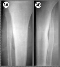
Rice. 3. Radiographs after surgery. The absence of a bone defect is visible on maternal fibula and the place from the depression on tibia. A - anteroposterior projection; B - lateral projection
The osteochondral exostosis was removed en bloc (Fig. 4 A, B).
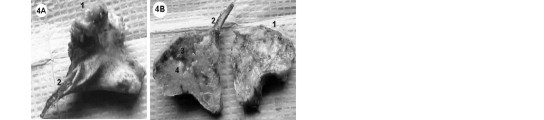
Rice. 4. Macroscopic view of osteochondral exostosis removed en bloc (A). Macroscopic view after sawing of osteochondral exostosis (B). 1- fibrous capsule, a cap-shaped cap made of hyaline cartilage, which functions as perichondrium; 2- wide base; 3- cortical and spongy bone; 4- hyaline cartilage, which in several areas grows towards the medullary component
The patient was discharged after the wound healed. At the time of discharge, the limb was not immobilized. No neurological disorders were noted. The patient walks independently without additional load.
During follow-up examinations, after 1, 6 and 12 months, he served in the armed forces.
As can be seen from the above examples, with the same treatment result, in the first case, where a wide resection was performed to create a bone defect, the rehabilitation period was extended. Additional fixation was required. In the second case, no additional fixation was used; the patient was activated after three days and discharged once the wound had healed.
Analysis of the above results indicates that in all cases, after surgical treatment, no relapses of the underlying disease were observed. No intraoperative complications or wound infections were noted in both groups.
Thus, as a result of the data presented, we came to the following conclusions:
- With a verified diagnosis of osteochondroma, there is no need for major surgical intervention with marginal bone resection within the healthy bone, which sharply prolongs the patient’s recovery time, especially when we're talking about about the lower extremities that are exposed to static loads.
- Extended marginal or parietal bone resection with removal root osteochondral exostosis leads to unjustified creation iatrogenic bone defect, which in some cases has to be filled with a graft. Full recovery bone structure in the area of the bone defect lasts up to 2 years and sometimes pathological fractures occur. This is an aggressive and unjustified approach. This result in the treatment of osteochondroma, taking into account the modern pace of life and the requirements of patients, it can hardly be considered satisfactory.
- Indications for surgical intervention for osteochondromas of the upper and lower limbs should be determined by the degree of secondary complications caused by exostoses, and not by the principle that the bone formation must be removed. When deciding on a possible surgical intervention The location of the osteochondroma and the likelihood of its fracture should also be taken into account.
- The results of our study show the effectiveness intact operations and make it possible to achieve similar therapeutic results with an incomparably quick return of the patient to a full life and, no less important, improving the quality of life of patients during the treatment period.
Literature
- Bandiera S., Bacchini P., Bertoni F. Bizarre parosteal osteochondromatous proliferation of bone, Skeletal Radiol., 1998; 27:154-156.
- Bell R.S. Musculoskeletal images: malignant transformation in familial osteochondromatosis? Can. J. Surg., 1999; 42:8.
- Dahlin D.C. and Unni K.K. Bone Tumors. General Aspects and Data on 8,542 Cases. Ed. 4, Springfield, Illinois, 1986.
- D'Ambrosia R. and Ferguson A.B. J. The formation of osteochondroms by epiphyseal cartilage transplantation, Clin. Orthop., 1968, 61: 103-115.
- Geirnaerdt M.J., Hogendoorn P.C., Bloem J.L., Taminiau A.H., van der Woude H.J. Cartialginous tumors: fast contrast-enhanced MR imaging, Radiology, 2000; 214:539-546.
- Harper G.D., Dicks-Mireaux C., Leiper A.D. Total body irradiation-induced osteochondromata, J. Pediatr. Orthop., 1998; 18:356-358
- James H. Beaty, Canale Terry: Operative Pediatric Orthopedics, 1991, p. 1098-1099.
- Kingsley R. Chin, F. Daniel Kharrazi, Bruce S. Miller, Henry J. Mankin, and Mark C. Gebhardt, Osteochondromas of the distal aspect of the tibia or fibula, J. Bone and Joint Surg., 2000, 82:1269 .
- Lovell W.W., Winter R.B. and Morrissey R.T. Pediatric Orthopedics, Third Edition, 1990, vol. 1, p. 342-343.
- Mark D. Murphey, James J. Choi, Mark J. Kransdorf Imaging of Osteochondroma: Variants and complications with radiologic-pathologic correlation, radiographics, 2000, 20: 1407-1434.
- Milgram J.W. The origins of osteochondromas and enchondromas. A histopathologic study, Clin. Orthop., 1983,74: 264-284.
- Mirra J.M. Bone Tumors. Clinical, Radiologic, and Pathologic Correlation. Vol. 2, Philadephia, 1989.
- Ozaki T., Hillmann A., Blasius S., Link T., Winkelmann W. Multicentric malignant transformation of multiple exostoses, Skeletal Radiol., 1998; 27:233-236.
- Resnick D., Kyriakos M., Grennway G.D. Osteochondroma. In: Resnick D. eds. Diagnosis of bone and joint disorders. 3rd ed., vol. 5, Philadelphia, 1995, p. 3725-3746.
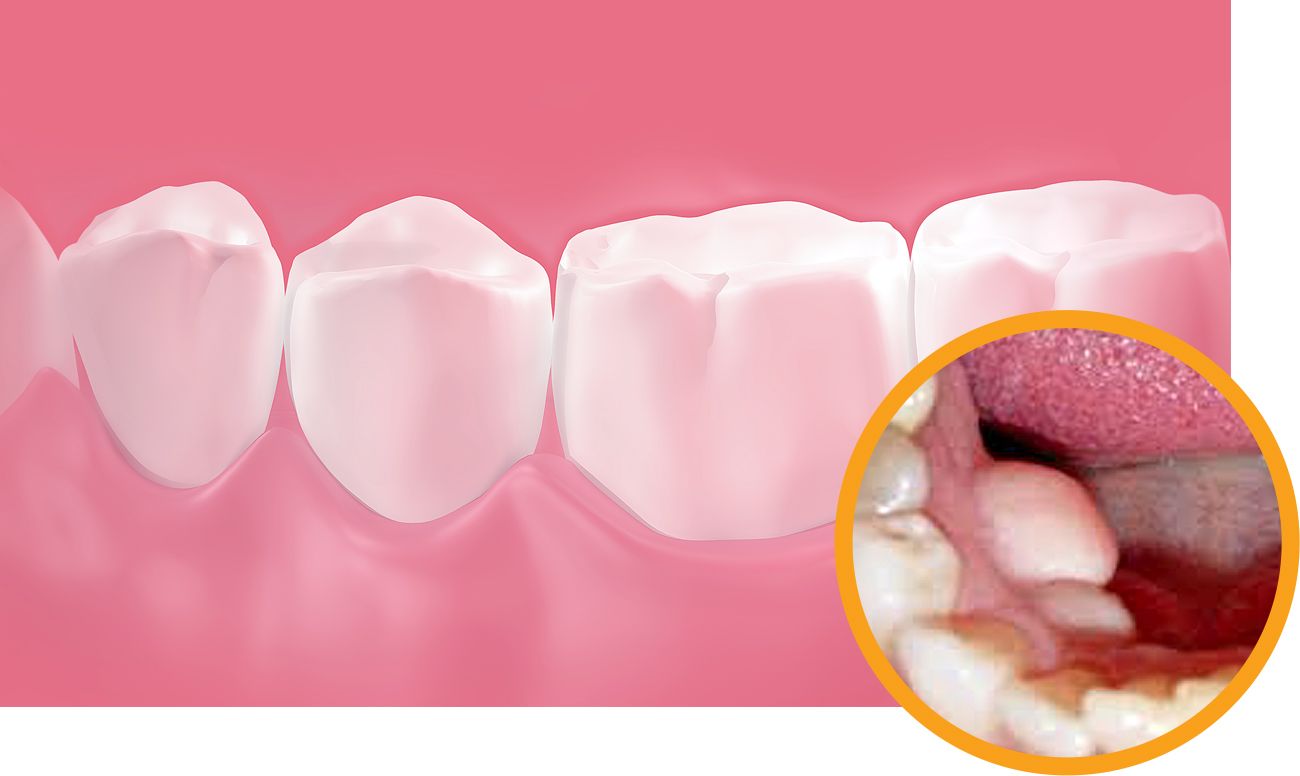
Exostosis is a disease accompanied by jaw anomalies, which manifests itself in the form bone growths and protrusions. Such protrusions often lead to additional pressure on adjacent teeth and bones, and also interfere with the wearing of dentures. This is due to the fact that when they put pressure on bone formation, a person develops severe pain and additional inconveniences appear while wearing the prosthesis.
Why do exostoses form?
As a rule, the formation of exostoses is explained by one of two factors - this or individual feature the structure of the human jaw, or the consequence of tooth extraction, in which the bone tissue was damaged or simply could not heal, as a result of which it shifted to the side. This displaced bone tissue may often not cause unnecessary inconvenience or affect the condition oral cavity and the functioning of the jaw apparatus, since a small growth is almost invisible and appears in the form of a small bump on the gums. But over time, the pressure on the bone and adjacent teeth may increase, and exostosis itself, being benign growth, can develop into a malignant tumor.
How is bone exostosis treated?
The only option for getting rid of the growth is surgery to remove the exostosis. After an X-ray examination, the attending physician receives all the necessary information about the growth and begins preparing the patient for surgery. Removal bone exostosis is happening as follows:
The time it takes to perform such an operation depends on the size of the exostosis and the complexity of the disease, but on average it lasts no more than two hours. In some cases, in addition to suturing, a special pressure bandage, which consolidates its result.
After surgery
After surgery to remove exostosis, doctors recommend extreme caution to avoid suture dehiscence. In this regard, certain temperature regime with drinks, take soft warm food, and also refrain from bad habits at least for a while.
Patients who strictly follow the instructions of their attending physician undergo rehabilitation period in about a week. But in some cases, wound healing and recovery after surgery can occur within a month. This is possible if the patient increased amount germs in the mouth or reduced immunity.
Some tumors in the mouth appear due to serious illnesses, and some may appear completely unexpectedly, for no apparent reason. Even if they are absolutely harmless, they will still have to be treated or removed, otherwise you will not be able to get rid of the discomfort. Such anomalies include exostosis.
What is exostosis and why does it occur?
Exostosis in dentistry is called growths - the process of growth of osteochondral or bone tissue, manifested in the form of protrusions on the surface of the palate or lower jaw. They can be attributed to benign neoplasms, they do not cause any pain.
The main problem is that exostosis gradually increases. This leads to increased discomfort, pressure on teeth and bones increases, and the risk of injury to the oral mucosa increases. With such neoplasms it is impossible to wear prostheses and place implants, and among them possible consequences– malocclusion and chin displacement.
On initial stage exostosis may be invisible and can only be detected by a doctor during a dental examination or x-ray. Large growths are easily felt by the tongue.
The reasons why exostosis appears are different:
- congenital jaw anomalies;
- bruises, fractures and other injuries;
- heredity;
- endocrine diseases;
- infectious diseases.
Exostosis often occurs after tooth extraction with complications. At serious damage bone tissue shift to the side, grow together incorrectly and bone development begins.
How to treat?
It is impossible to cure exostoses on the gums at home, without surgical intervention it won't work out. The operation must be performed by a dental surgeon. Of course, the procedure has its limitations: contraindications include diabetes mellitus, problems with endocrine system and adrenal glands, poor clotting blood.
If the growth on the jaw is very small and does not cause inconvenience, with surgical removal you don't have to rush. However, someday you still have to do this. An overgrown exostosis will interfere not only with the tongue, but also adjacent teeth, which threatens deformation. And if you need a prosthesis, it is the growths on the jaw that will become the main obstacle to installing implants.
During removal, the specialist will numb the surrounding tissue growths (usually used local anesthesia) and make a small incision on the gum. The neoplasm is cut down and smoothed, after which sutures are placed on the jaw mucosa. Removal process bone formation on the gums can last several hours, the duration of the procedure will depend on the size of the exostosis and its location.
The cost of the operation will depend on the chosen dentistry, the amount of exostosis, the number of problem areas and the chosen method of anesthesia.
What to do after surgery
The first days after surgery, you need to carefully monitor the condition of the stitches and do everything to prevent them from coming apart. It is necessary to temporarily avoid solid and tough foods. Very hot or cold drinks, alcohol and cigarettes will slow down the healing process. It is recommended to reduce physical activity and avoid stress.
Minor swelling and pain may occur, recovery period You can use painkillers and decongestants. Of course special attention You should pay attention to oral hygiene: it is better to consult with your dentist about what products are best to rinse your mouth to prevent bacterial infection.
If the operation to remove exostosis of the jaw was carried out efficiently and the patient followed all the recommendations, there should be no complications. In case of prolonged pain, fever and swelling that does not subside, you should immediately consult a doctor. In addition, you should not prescribe treatment for yourself - this can only worsen the rehabilitation process.


