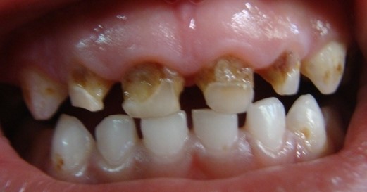
Caries is one of the most common dental disease on our planet. Its presence on the surface of the teeth requires mandatory medical intervention in order to prevent their further destruction. And the caries classification system will help you choose a treatment method for a particular clinical case.
Black's classification of carious formations on the surface of teeth was proposed in 1896 in order to determine treatment standards for each individual clinical case.
It included five classes, each of which had its own method of preparing and filling teeth. After the sixth grade was added to the classification, it remained unchanged to this day.
Class I
The first class includes carious lesions of the pits, fissures and natural depressions of the chewing, palatal or buccal surfaces of the teeth - the so-called fissure caries.
Class II
The second class includes caries of the contact surfaces of molars and premolars.
Class III
The third class includes caries contact surface incisors and canines, without affecting the integrity of their cutting edges.

Class IV
The next stage is a more intense damage to the incisors and canines, violating the integrity of their cutting edge.

Class V
The fifth class includes damage to the vestibular surface of all groups of teeth - cervical caries.
Class VI
The sixth class includes caries located on the tubercles of the molars and the cutting edges of the incisors and canines.

Classification of caries according to ICD-10 (WHO)
The ICD-10 (World Health Organization) classification is as follows:
- dental enamel caries;
- dentin caries;
- cement caries;
- caries that has stopped due to exposure to hygienic and preventive procedures;
- odontoclasia, characterized by resorption of the roots of primary teeth;
- other caries;
- unspecified caries.
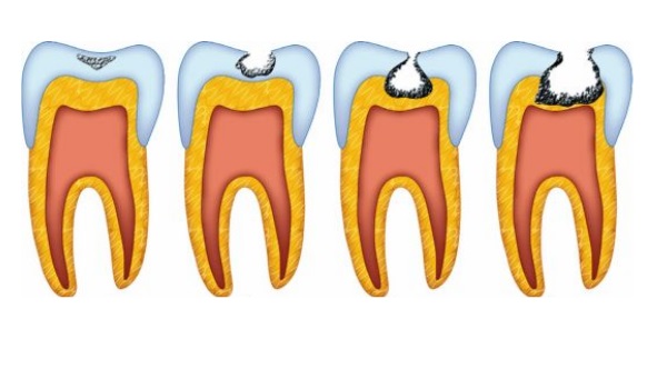
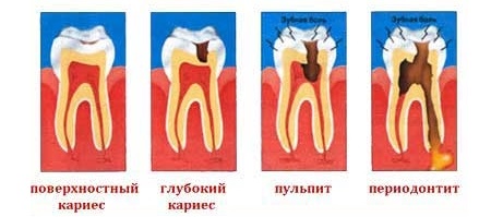
According to the depth of the lesion
Based on the depth of damage, caries is divided into several stages.
These include:
- initial caries;
- superficial caries;
- average caries;
- deep caries.
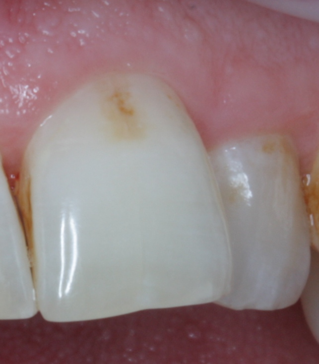
Initial caries
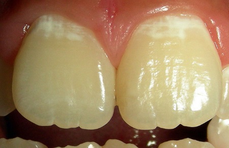
The initial stage of development of the disease begins with the formation of a white or dark spot on the surface of the tooth. At the same time, the enamel remains smooth to the touch, since it has not yet reached the point of anatomical destruction.
There is no toothache at this stage, and treatment is carried out with minimal intervention in its structure.
The resulting stain is removed using dental equipment and the teeth are remineralized to prevent further development carious process.
The next stage in the development of caries is the destruction of the upper layers of enamel with the appearance of a reaction to sudden change food and water temperatures, and sour or spicy foods.
The smoothness of the tooth surface is disrupted and it becomes rough.
Treatment at this stage includes resurfacing of the affected area followed by remineralization. Applies also traditional treatment with preparation and filling.

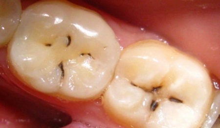
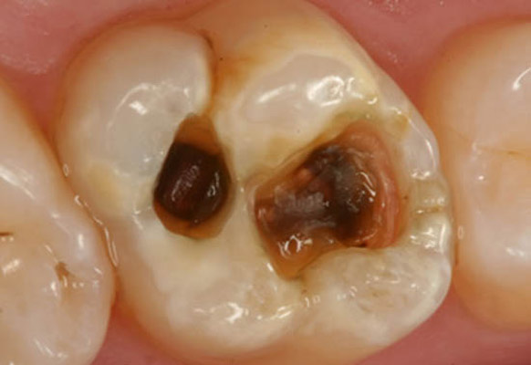
Medium caries means the destruction of the enamel layer of the tooth with the appearance of periodic or permanent pain. This is due to the fact that the pathogenic process has affected the upper layers of dentin.
Average caries requires mandatory medical intervention, which involves removing the affected area and then restoring it with filling material.

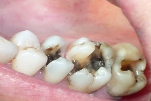
Deep caries is characterized by extensive damage to the internal tissues of the teeth, affecting most of the dentin.
Ignoring this process and failure to carry out treatment can lead to damage to the pulp with subsequent complication of the disease pulpitis and/or periodontitis. Therefore, the affected area must be removed for subsequent installation of a filling.
Video: types of caries
According to the presence of complications
Based on the presence of complications, caries is divided into complicated and uncomplicated.
Uncomplicated
The uncomplicated caries process includes a typical carious process, including its various stages (superficial, medium, deep).
Complicated
Complicated caries includes a disease accompanied by the development of concomitant inflammatory processes. Most often, this is a consequence of late consultation with a doctor or insufficient treatment.
By degree of activity
To assess the degree of disease activity, the Vinogradova classification is used, based on the division of caries into compensated, subcompensated and decompensated.
Compensated
Compensated caries is characterized by a sluggish or non-progressive process. Damage to the surface of the teeth is insignificant and does not cause any discomfort in the patient.
With regular hygiene procedures, as well as holding special preventive measures it is possible to stop the development of the disease at its initial stages.
Subcompensated
Subcompensated caries is characterized by an average rate of progression, at which it can go unnoticed and not cause concern to the patient at all.
Decompensated
Decompensated caries is characterized by intensive development and progression, accompanied by such acute pain that it affects the patient’s ability to work. Because of this, the disease is often called acute caries.
It requires immediate medical procedures, since otherwise the process may spread to third-party teeth with the subsequent addition of pulpitis and periodontitis.
According to the nature of the flow
According to the nature of the course, caries is divided into acute, chronic, acute and recurrent.
- Acute caries characterized by the appearance of signs of dental damage within just a few weeks.
- Chronic caries develops over a longer period of time. At the same time, the affected tissues have time to become stained with plaque and food dyes, acquiring colors from yellow to dark brown.
- Acute or blooming caries characterized by multiple lesions of dental tissue within a fairly short time. This phenomenon is often observed in children with low immunity, as well as in adults after removal salivary glands accompanied by dry mouth.
- Recurrent and secondary caries is a consequence of a number of provoking factors. These include damage or weakening of tooth enamel, failure to comply with personal hygiene rules, as well as decreased immunity due to any diseases of the body.
According to the intensity of the process
According to the intensity of the process, the disease is divided into single and multiple caries.
In the first case, one tooth is involved in the process, and in the second - several teeth at the same time. Defeat large quantity teeth in a short period of time is called generalized caries.
By process localization
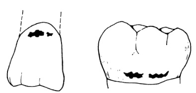
According to the localization of the process, caries is divided into fissure, interdental, cervical, circular and hidden.
- Fissure or occlusal caries characterized by the development of lesions in the natural recesses of the chewing surface of the teeth.
- Interdental or proximal caries develops on the contact surfaces of teeth, and for a long time may not be visualized. This is due to the specifics of the development of the disease: affecting the surface of the tooth, caries develops towards its center, while the cavity itself is often covered by a preserved layer of enamel. You can find it using x-ray or by dark areas showing through the teeth.
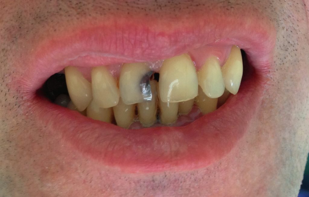
- Cervical or cervical caries develops in areas of the teeth located between their crown and root closer to the gum - on the neck. It is a consequence of insufficient oral hygiene.
- Circular or ring caries characterized by circumferential damage to the tooth surface. The appearance of the disease resembles a yellow or brown belt around the neck of the teeth, with more than half clinical cases falls on children.
- Hidden caries characterized by damage to areas that are difficult to see, such as dental crevices.
According to the primacy of development
Based on the priority of development, caries is divided into primary and secondary.
Primary caries develops either on an intact tooth or on an area that has not previously been treated.

Secondary caries is recurrent because it appears on the treated sites, that is, where a filling was previously installed. Due to the fact that the location of the disease is often the area located under a filling or dental crown, it is called internal caries.
Video: why fillings need to be replaced
Classification in children
The principles of classification of caries in children are practically no different from adults. The only difference is the division of its parameters into caries permanent teeth and caries of primary teeth.
In the latter case, the picture of the lesion is of the same nature as in adults, but due to the temporary purpose of baby teeth, treatment is carried out somewhat differently.
According to changes in the hard tissues of the tooth and clinical manifestations, several types have been created classification of dental caries , they are based on various features.
Caries is one of the most well-known diseases affecting the hard tissues of the tooth. The process of development of the disease is accompanied by thinning of the enamel, softening of dentin and the formation of a carious cavity. Speaking about dental caries, it is impossible to limit ourselves to just one classification that would fully satisfy the requirements of specialists. Therefore, the existence of several classifications of the disease is quite justified.
Classification of caries according to Black
The greatest recognition among dentists today is the Black classification of caries, which reflects the depth of the process, as well as the location carious cavities.
1)
. First class
(superficial caries
). The cavities are located in the area of natural depressions and fissures. The defeat is superficial;
2)
. Second class
(weak caries
). The process develops on the contact surface of the lateral teeth;
3)
. Third class
(caries medium degree
). Carious lesions affect the contact surface of the canines and incisors;
4)
. Fourth grade
(severe form of caries
). Advanced stage of moderate caries. Carious lesions move onto the dentin at the incisal angle;
5)
. Fifth grade
(very severe caries
). The gingival margin of the lateral or front teeth suffers. Radical caries develops;
6)
. Sixth grade
(atypical caries
). Destruction of the cutting edge is observed.
Classification of the disease according to ICD-10 | WHO
Depending on the nature of the changes occurring in the hard tissues of the tooth, as well as clinical manifestations, several ways have been created classify dental caries .
ICD caries presupposes the presence different signs at the core. By WHO classification caries stands out in a separate group.
 ICD-10 suggests dividing caries into the following classes:
K02.0 Enamel caries chalk spot stage (initial caries)
K02.1 Dentin caries
K02.2 Cement caries
K02.3 Suspended dental caries
K.02.3 Odontoclasia
Pediatric melanodentia
Melanodontoclasia
K02.8 Other dental caries
K02.9 Dental caries, unspecified
Classification of caries according to ICD 10 on at the moment is one of the most popular. Among its advantages we can attribute the fact that sub-categories appeared in it in the form of suspended caries or cement caries.
Classification of the carious process by the depth of the lesion | MMSI
 Dentists consider this classification of caries to be the most convenient. Therefore, it has become widespread in the domestic space. Experts distinguish forms of the disease related to uncomplicated and complicated course of the disease:
 1. Spot stage – initial stage when white streaks appear on the enamel or dark spots, but it itself is smooth to the touch and is not yet subject to destruction. Toothache at this stage of the stain does not bother the patient;
2. Superficial caries – the second stage of the carious process. Tooth enamel continues to deteriorate, but caries does not yet extend beyond the enamel layer. Dentin is not damaged, however, toothache of a periodic nature may already manifest itself. The reaction of the tooth to cold and hot, sour or sweet is noticeable. A carious stain on the tooth surface is rough to the touch;
3. Moderate caries , when the carious lesion has passed the enamel layer and affected the upper layers of dentin. The pain intensifies and is constant;
4. Deep caries , in which only a thin layer of dentin is preserved. At this stage, the dental tissue is severely damaged. Lack of proper tooth treatment at this stage causes pulp damage and periodontitis.
Classification according to the presence of complications
This classification involves distinguishing two types of caries:
- complicated accompanied by concomitant inflammatory processes. This form of the disease occurs when a doctor is not consulted in a timely manner or due to the lack of proper treatment;
- uncomplicated – a typically occurring process, which presupposes the presence of its individual stages (superficial, middle, etc.).
 Types of caries by degree of activity:
1. Compensated caries , characterized by the absence of obvious progress in the carious process. The teeth are slightly affected, which does not cause discomfort to the patient;
2. Subcompensated , characterized by an average rate of development;
3. Decompensated , which is characterized by intense flow. At this stage it is diagnosed sharp pain in the tooth.
This classification is based on calculating the caries intensity index, which is defined as the sum of carious, filled and extracted teeth (CP) in one child. If there are both milk teeth and permanent teeth in the oral cavity, then the amount is calculated for them separately (KPU + KP). Extracted baby teeth are not counted.
How quickly does the carious process develop?
In this case, the classification is a composition of the following four categories :
- acute caries . Signs of dental damage appear within a matter of weeks;
- chronic caries , developing over a longer period of time. Affected tissues take on a yellowish or dark brown color, stained with plaque and food coloring;
- blooming caries , which entails multiple lesions of dental tissue. The carious process progresses over a short time;
- secondary caries , developing under a previously installed filling as a result of weakening of tooth enamel, neglect of the rules of oral hygiene, and decreased immunity of the body.
Classification of the disease according to the intensity of the process
This classification assumes the presence of:
single caries . In this case, only one tooth is affected;
multiple (systemic) caries . This form of the disease affects five or more teeth in children, six or more in adults.
Patients with such a diagnosis most often include those who suffer from acute infectious diseases, cardiovascular diseases, respiratory system. Among children suffering from multiple caries, there are those who have recovered from the disease. chronic tonsillitis, scarlet fever .
Classification by process localization
 -
fissure caries
, in which the natural recesses of the surface of the teeth are affected;
-
fissure caries
, in which the natural recesses of the surface of the teeth are affected; - interdental caries process , developing on the contact surface of the tooth. Long time the disease may not be diagnosed due to specific shape development of the disease: caries, in the process of damage to the tooth surface, develops towards the center of the tooth, and the cavity itself is covered with healthy enamel layers;
- cervical caries , which is localized between the root and crown of the tooth, in the area adjacent to the gum. The reason for the development of the process is poor hygiene oral cavity;
- ring caries , affecting the circumferential surface of the tooth. Outwardly it looks like a yellowish or brown belt on the neck;
- hidden carious process , developing in a difficult-to-see area - the dental crevice.
Classification according to the primacy of development
 It is not difficult to guess that this classification divides caries into:
- primary , which affects either healthy tooth, or an area that has not previously been treated;
- secondary , which is recurrent in nature, because it develops in previously healed areas.
Sometimes this type of carious process is called internal: the disease is often localized in the area under the filling or crown.
Clinical classification of dental caries
- Acute caries . It is typical for him rapid development destructive changes in the hard tissues of the tooth, the rapid transition of uncomplicated caries to complicated ones. The affected tissues are soft, slightly pigmented (light yellow, grayish-white), moist, and can be easily removed with an excavator.
- Chronic caries characterized as a slow process (several years). The spread of the carious process (cavity) is mainly in the planar direction. The altered tissues are hard, pigmented, brown or dark brown in color.
- There are also other forms of caries , for example, “acute”, “blooming caries”.
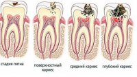
In our country, this classification is most widely used. It takes into account the depth of the lesion ,
 which is very convenient for practical activities dentist
 - Stage of carious spot – focal demineralization of the hard tissues of the tooth is observed, and it can proceed intensively ( white spot) or slowly (brown spot).
- Superficial caries – at this stage a carious cavity appears within the enamel.
- Average caries – at this stage, the carious defect is located within the surface layer of dentin (mantle dentin).
- Deep caries - in this case pathological process reaches the deep layers of dentin (peripulpal dentin).
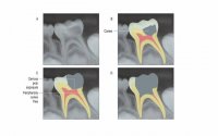
IN clinical practice The terms “secondary caries” and “recurrent caries” are also used; let’s take a closer look at what they are:
1)
Secondary caries
– these are all new carious lesions that develop next to the filling in a previously treated tooth. Secondary caries has all the histological characteristics of a carious lesion. The reason for its occurrence is a violation of the marginal seal between the filling and hard tissues tooth, microorganisms from the oral cavity penetrate into the resulting gap and optimal conditions are created for the formation of a carious defect along the edge of the filling in the enamel or dentin.
2)
Recurrence of caries
– this is the resumption or progression of the pathological process if the carious lesion was not completely removed during previous treatment. Recurrence of caries is more often found under a filling when X-ray examination or along the edge of the filling.
 quite a lot and they are all largely repeated. It is important for the doctor to correctly determine the main parameters: the depth of the lesion, the nature of the process, and to identify the main cause of the defects.
In some cases this will be unsatisfactory oral hygiene, in others - bad habits, thirdly, crowded teeth or congenital disorders in the structure of enamel and dentin.
In the International Statistical Classification of Diseases and Related Health Problems of the World Health Organization, Tenth Revision (ICD-10):
K02.0 Enamel caries
Stage of “white (chalky) spot” [initial caries]
K02.1 Dentin caries
K02.2 Cement caries
K02.3 Suspended dental caries
K02.4 Odontoclasia
K02.8 Other dental caries
K02.9 Dental caries, unspecified
General approaches to the diagnosis and treatment of dental caries:
Diagnosis of dental caries is made by collecting anamnesis, clinical examination And additional methods examinations. The main task in diagnosis is to determine the stage of development of the carious process and select the appropriate treatment method. During diagnosis, the localization of caries and the degree of destruction of the crown part of the tooth are established. Depending on the diagnosis, a treatment method is chosen.
The principles of treating patients with dental caries provide for the simultaneous solution of several problems:
Elimination of factors determining the demineralization process;
Prevention of further development of the pathological carious process;
Saving and Restoring anatomical shape a tooth affected by caries and the functional ability of the entire dental system;
Prevention of the development of pathological processes and complications;
Improving the quality of life of patients. Treatment for caries may include:
Elimination of microorganisms from the surface of teeth;
Remineralizing therapy at the “white (chalky) spot” stage;
Fluoridation of hard dental tissues for suspended caries;
Preservation of healthy hard dental tissues whenever possible, excision of pathologically altered tissues with subsequent restoration of the tooth crown;
At taking anamnesis determine the presence of complaints of pain from chemical and temperature stimuli, allergic history, availability somatic diseases. Targeted identification of complaints of pain and discomfort in the area of a specific tooth, complaints of food getting stuck, patient satisfaction appearance tooth, the timing of the onset of complaints, when the patient noticed the appearance of discomfort. Determine whether the patient is carrying out proper hygiene care for the oral cavity, the patient’s profession, regions of his birth and residence (endemic areas of fluorosis).
Caries is a pathological process of gradual destruction of hard tooth tissues with the formation of carious cavities due to demineralization.
The International Classification of Dental Diseases (ICD-10) divides dental caries into the following categories:
| K02.0 | K02.1 | K02.2 | |
| Enamel caries | Dentin caries | Cement caries | |
| K02.3 | K02.4 | K02.8 | K02.9 |
| Suspended caries | Odontoclasia | Other specified caries | Unspecified dental caries |
- Tooth enamel caries- a reversible process that can be easily eliminated. It is considered the initial stage of caries. Damage affects surface layer enamels. Another name is caries in the spot stage.
- Dentin tissue caries begins due to deficiency minerals as a result of damage to tooth enamel. Dentin has lower strength characteristics compared to enamel. Therefore, the development of caries at this stage proceeds faster. This is an irreversible process that must be treated.
- Cement caries develops as a result of inflammatory processes in the area of the tooth root. It is rare and requires immediate treatment. Leads to tooth loss and the occurrence of pulpitis or periodontitis.
- Suspended course of caries often observed after remineralization procedures. Indicates stopping further damage to dental tissue.
- Odontoclasia– a pathological process that is characterized by damage to the enamel and the formation of carious cavities.
- Other types of caries– other forms of carious lesions not presented in the typology.
Classification Features
Classification of caries according to ICD-10 is successfully used in modern practice dentistry, but, unfortunately, has a number of significant disadvantages. The division of caries according to the location in the dental tissues provides incomplete information about the course of the disease. For clarification clinical picture There are a number of other typologies that are used to study the depth of damage, the course and intensity of the disease.
Causes of caries
Caries has an infectious nature. It is believed that the destructive effect on tooth enamel and other dental tissues have streptococcal bacteria: Streptococcus mutans And Streptococcus sanguis.
But it should be noted that in the microflora of the oral cavity these bacteria can exist in small quantities healthy person and not cause pathological processes. The activity and harmfulness of bacteria largely depends on the state of the human body as a whole and various negative factors.
Risk factors for developing caries:
- Domestic(endogenous):
- hypovitaminosis;
- decreased immunity;
- the presence of gastrointestinal pathologies and other chronic diseases;
- hereditary predisposition;
- low level of caries resistance of tooth enamel;
- stress.
- External(exogenous):
- unbalanced diet with a predominance of carbohydrates and sugar;
- poor oral hygiene, dental plaque;
- irregular use of fluoride;
- the presence of dentures, braces on the teeth, or uneven tooth growth (having hard-to-reach places for cleaning promotes the growth of bacteria).
The stages of caries are distinguished according to the course of the disease:
- Initial stage.
- Superficial caries.
- Average caries.
- Deep.
Initial stage caries development means pathological formations stains on the surface of tooth enamel. Easily eliminated with the help of preventive measures.
Signs:
- The formation of a white matte spot on the tooth surface, indicating a focus of demineralization.
- Smooth yellow-brown spots are found.
At the stage superficial caries The tooth enamel is affected and irreversible changes begin.
Signs:
- The spot is distinct, rough;
- Painful sensations after eating sweet, sour, hot or cold food;
- The beginning of the appearance of a carious cavity.
Stage of middle caries characterized by the localization of pathological processes already in dentin. The lesion penetrates deep into the tooth.
Signs:
- Shallow carious lesions of dentin;
- Painful reaction to thermal and chemical irritants;
- Inflammation of the gums.
Deep caries accompanied by extensive carious lesions that affect the cementum tissue of the tooth. At this stage, the patient clearly feels pain, which may be permanent. It requires immediate treatment, as it leads to tooth loss and the development of other oral diseases (pulpitis).
Signs:
- Deep lesions reaching the tooth root;
- Softened tooth tissues are visible in the hole, food debris accumulates;
- Pronounced pain syndrome;
- Soreness and swelling of the gums.
Treatment of caries
Treatment of caries on chalk spot stages includes saturation of the tooth surface with calcium and fluoride, as well as general prevention development of carious processes. Medium and superficial caries It is treated by eliminating the affected area using a drill and further filling.
Deep caries are treated in several stages using additional medications to disinfect and mineralize the carious cavity. Filling is performed after removing the altered tooth tissue.
Other classifications of caries
Caries has several dozen classifications based on certain characteristics. They help to more accurately identify the nature and causes of the disease.
For example, according to the location of the lesion, caries is divided into:
- Fissure– the focus of the disease on the fissure of the tooth (chewing surface).
- Contact– the lesion is located between the contacting teeth.
- Cervical- development pathological changes in the area of the tooth root, closer to the gum.

Carious lesions are:
- Single;
- Multiple;
- System.
The course of the disease can be of different types:
- Fast flowing;
- Slow;
- Stabilized.
Separation according to the primacy of the disease:
- Primary – disease of a healthy tooth;
- Secondary (relapse) – the development of caries in the area of the filling due to improper installation or processes of destruction of the material.
Prevention of caries
Caries develops as a result of exposure to pathogenic microflora, which occurs due to various reasons. The main goal is to eliminate this cause, which provokes the destruction of dental tissue.
To do this, you should know and adhere to certain rules for caries prevention:
- Careful oral hygiene. Regular brushing of teeth and tongue will prevent the formation of harmful bacteria; rinsing your mouth is also important by special means, which help improve microflora and have antimicrobial effect. In addition, many rinses are fortified with calcium and fluoride, which helps restore mineral balance.
- Professional teeth cleaning to remove plaque and tartar. Brushing with a toothbrush will not completely remove plaque; a lot of it accumulates in hard-to-reach places. These are potential sites for tooth decay to develop. To avoid the development of the disease, you need to carry out professional cleaning teeth by chemical or mechanical method.
- Balanced diet. Predominance in nutrition natural products, adequate protein intake and avoidance of carbohydrate foods with high content starch, as well as minimal sugar consumption contribute not only to the prevention of caries, but also other diseases.
- Correction incorrect position dentition. If the dentition is formed incorrectly, areas appear where the teeth touch and overlap each other. Bacteria accumulate in hard-to-reach areas and cause caries. The same applies to incorrectly installed orthodontic structures, prostheses. The doctor will help solve this problem.
- Regular visits to the dentist. In order to prevent the development of caries, or notice its manifestation on initial stage you need to visit a specialist several times a year. At the first stage, caries is asymptomatic and can only be diagnosed by a dentist.
- Strengthening the immune system. Weakening protective forces the body provokes many diseases. The microflora of the oral cavity is disrupted, and destructive processes occur. Strengthening the body prevents the development of microorganisms of the genus staphylococcus, which reduces the risk of caries.
ICD 10 caries is international classification diseases according to the category of caries, which is accepted World Organization health care in the 10th edition. Experts have created many systems for separating carious lesions depending on the prevalence of the pathological process, its localization and clinical course.
According to modern medical concepts, dental caries according to ICD 10 is considered infectious disease. In this regard, the key cause of carious lesions is bacterial contamination of dental surfaces.

The following factors contribute to the formation of a carious defect:
Nutritional Features
Numerous clinical observations indicate that overuse sweets causes multiple dental caries.
It is very important to note that the destructive effect of carbohydrates occurs when sucrose comes into close contact with the enamel surface. In such cases, the formation of a carious cavity still requires the presence of pathological microorganisms and an insufficient level of oral hygiene.

Such gross violations food composition, such as insufficient levels of proteins and vitamins, also serves as an indirect cause of carious lesions of hard tooth tissues. This also affects the quality properties of saliva, which becomes more viscous and loses its protective properties.
One of the decisive cariogenic causes is the consistency of the food consumed. Soft foods create conditions for the formation of plaque and the formation of enamel defects.
Past diseases
Some scientists have expressed the opinion that some chronic diseases(tuberculosis, diabetes mellitus etc.) have a direct impact on the severity of the carious process. This occurs due to a disruption in the blood supply to the dental pulp, which is responsible for the mineralization and strength of the enamel.
The most pronounced toxic effect on dental tissues transferred systemic diseases have during the period of change of milk teeth. At this time, the patient’s saliva composition changes significantly, which loses its protective properties.
External risk factors
Among the external cariogenic factors, ionizing effects should be noted. So, 6-8 weeks after completion of the course radiation therapy a person has multiple chalky spots on the surfaces of teeth. Over time, these pigmentations turn into carious defects.
Very often caries, this form of the disease ends complete destruction crowns As a result, the patient requires orthopedic treatment. The pathological process is aggravated by the forced intake of soft carbohydrate foods.
IN dental practice Radiation damage to enamel and dentin is commonly called radiation caries.
Microbial factor
The presence of carbohydrates in the oral cavity and pathogenic bacteria is prerequisite caries development. Based on the results of numerous scientific research Experts have found that demineralization and destruction of enamel are caused by organic acids produced by bacteria.
An increase in the activity of pathological microflora is facilitated by a decrease in the acidity of the oral fluid, the deposition of dental plaque and frequent consumption of carbohydrates. That is why the instructions for providing dental care The population requires priority cleansing of the oral cavity from bacterial plaque.

Main types of dental caries
The standard classification of caries according to ICD 10 includes the following categories:
- K 02.0 Initial caries, which is located within the enamel;
- K 021 Medium and deep caries or carious lesions of dentin;
- K 02.2 Root caries;
- By 02.3 Caries at the stage of suspension of the pathological process;
- K 02.8 Other variants of carious lesions;
- By 02.9 Unrefined caries.
In his daily work, a dentist, as a rule, conventionally divides the disease according to topographical criteria.
Thus, the classification of caries according to the ICD includes:
| Stage of the carious process | Appearance of the affected tooth |
| Carious spot |
|
| Superficial caries |
|
| Average caries |
|
| Deep caries |
|

In dentistry, there is a classification of dental caries according to ICD 10, which takes into account the nature of the clinical course of the disease.
As a result, carious lesions occur in the following forms:
- Acute process. Caries spreads to enamel and dentin within 1-2 weeks. In this case, the carious cavity is filled with softened dentin and food debris.
- Chronic process. For a slowly occurring carious lesion, the absence of patient complaints and the formation of a pigmented defect is considered typical.
Experts also identify two additional forms of caries:
- secondary caries, when the pathological process is located in the gap between the filling and the tooth surface;
- relapse in the form of repeated carious lesions, which develops due to insufficient primary removal of necrotic masses.
In dental institutions, the generally accepted classification of caries according to Black and ICD, which includes the following classes:
- Carious cavities located in the natural cavities of the teeth (fissures).
- Demineralization of enamel and dentin on the lateral surfaces of small and great molars.
- Contact caries of incisors and canines while maintaining the integrity of their cutting edges.
- Caries on the contact sides of the anterior group of teeth with destruction of the cutting edges.
- Cervical caries of the upper and lower dentition.
We recommend reading
 Degas E. “Blue Dancers. Essay based on the painting by Edgar Degas “Blue Dancers Dancers in Blue”
 We reflect in the RSV reimbursement of social insurance expenses for the last year Appendix 2 line 090 of the calculation of insurance premiums
 Olesya Malibu: Russian clone of Pamela Anderson
 The problem of human spirituality